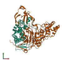
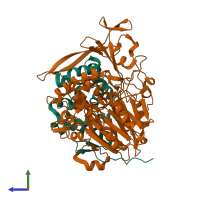
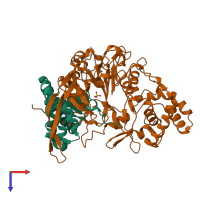
2ae4

X-ray diffraction
2.3Å resolution

Glutaryl 7-Aminocephalosporanic Acid Acylase: mutational study of activation mechanism

Released:

Function and Biology Details

Reaction catalysed:
(7R)-7-(4-carboxybutanamido)cephalosporanate + H(2)O = (7R)-7-aminocephalosporanate + glutarate
Biochemical function:
Biological process:
Cellular component:
  • not assigned

Structure analysis Details

Assembly composition:
hetero dimer (preferred)
PDBe Complex ID:
PDB-CPX-139683 (preferred)
Entry contents:
2 distinct polypeptide molecules
Macromolecules (2 distinct):
Glutaryl-7-aminocephalosporanic-acid acylase subunit alpha Chain: A
Molecule details ›
Chain: A
Length: 166 amino acids
Theoretical weight: 18.2 KDa
Source organism: Pseudomonas sp. GK16
Expression system: Escherichia coli
UniProt:
  • Canonical: P07662 (Residues: 30-195; Coverage: 24%)
Structure domains: Penicillin Amidohydrolase, domain 1
Glutaryl-7-aminocephalosporanic-acid acylase subunit beta Chain: B
Molecule details ›
Chain: B
Length: 528 amino acids
Theoretical weight: 59.17 KDa
Source organism: Pseudomonas sp. GK16
Expression system: Escherichia coli
UniProt:
  • Canonical: P07662 (Residues: 199-720; Coverage: 76%)
Sequence domains: Penicillin amidase
Structure domains:

Ligands and Environments

2 bound ligands:
No modified residues

Experiments and Validation Details

Entry percentile scores
X-ray source: PAL/PLS BEAMLINE 4A
Spacegroup: P41212
Unit cell:
a: 73.841Å b: 73.841Å c: 384.014Å
α: 90° β: 90° γ: 90°
R-values:
R R work R free
0.204 0.204 0.24
Expression system: Escherichia coli