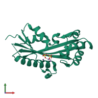
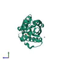
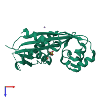
2amh

X-ray diffraction
2Å resolution

Crystal Structure of Maf-like Protein Tbru21784AAA from T.brucei

Released:
Source organism: Trypanosoma brucei
Entry authors: Caruthers J, Merritt E, Structural Genomics of Pathogenic Protozoa Consortium (SGPP)

Function and Biology Details

Biological process:
  • not assigned
Cellular component:
  • not assigned

Structure analysis Details

Assembly composition:
monomeric (preferred)
PDBe Complex ID:
PDB-CPX-162624 (preferred)
Entry contents:
1 distinct polypeptide molecule
Macromolecule:
septum formation protein MAF homologue, putative Chain: A
Molecule details ›
Chain: A
Length: 207 amino acids
Theoretical weight: 22.64 KDa
Source organism: Trypanosoma brucei
Expression system: Escherichia coli
Structure domains: Maf protein

Ligands and Environments

2 bound ligands:
No modified residues

Experiments and Validation Details

Entry percentile scores
X-ray source: ALS BEAMLINE 8.2.1, SSRL BEAMLINE BL1-5
Spacegroup: I4
Unit cell:
a: 97.068Å b: 97.068Å c: 48.894Å
α: 90° β: 90° γ: 90°
R-values:
R R work R free
0.199 0.197 0.231
Expression system: Escherichia coli