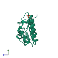
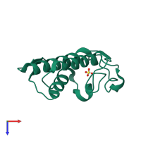
2aoz

X-ray diffraction
2.08Å resolution

Crystal structure of the myotoxin-II from Atropoides nummifer venom

Released:
Source organism: Atropoides nummifer
Primary publication:
Structure of myotoxin II, a catalytically inactive Lys49 phospholipase A2 homologue from Atropoides nummifer venom.
Acta Crystallogr Sect F Struct Biol Cryst Commun 62 423-6 (2006)
PMID: 16682766

Function and Biology Details

Structure analysis Details

Assembly composition:
monomeric (preferred)
PDBe Complex ID:
PDB-CPX-160655 (preferred)
Entry contents:
1 distinct polypeptide molecule
Macromolecule:
Basic phospholipase A2 homolog Chain: A
Molecule details ›
Chain: A
Length: 121 amino acids
Theoretical weight: 13.73 KDa
Source organism: Atropoides nummifer
UniProt:
  • Canonical: P82950 (Residues: 1-121; Coverage: 100%)
Sequence domains: Phospholipase A2
Structure domains: Phospholipase A2 domain

Ligands and Environments

1 bound ligand:
No modified residues

Experiments and Validation Details

Entry percentile scores
X-ray source: LNLS BEAMLINE D03B-MX1
Spacegroup: P43212
Unit cell:
a: 68.939Å b: 68.939Å c: 64.069Å
α: 90° β: 90° γ: 90°
R-values:
R R work R free
0.232 0.219 0.275