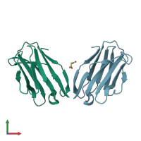
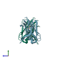
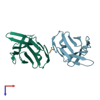
2apt

X-ray diffraction
2Å resolution

Crystal Structure of the G17E/S54N/K66E/Q72H/E80V/L81S/T87S/G96V variant of the murine T cell receptor V beta 8.2 domain

Released:
Source organism: Rattus norvegicus
Primary publication:
The nitty-gritty of protein interactions.
Structure 13 1737-8 (2005)
PMID: 16338399

Function and Biology Details

Structure analysis Details

Assembly composition:
monomeric (preferred)
Assembly name:
PDBe Complex ID:
PDB-CPX-106854 (preferred)
Entry contents:
1 distinct polypeptide molecule
Macromolecule:
Ig-like domain-containing protein Chains: A, B
Molecule details ›
Chains: A, B
Length: 112 amino acids
Theoretical weight: 12.16 KDa
Source organism: Rattus norvegicus
Expression system: Escherichia coli
UniProt:
  • Canonical: A2NAI0 (Residues: 1-94; Coverage: 99%)
Sequence domains: Immunoglobulin V-set domain
Structure domains: Immunoglobulins

Ligands and Environments

1 bound ligand:
No modified residues

Experiments and Validation Details

Entry percentile scores
X-ray source: RIGAKU RU200
Spacegroup: C2
Unit cell:
a: 79.423Å b: 116.295Å c: 34.769Å
α: 90° β: 110.17° γ: 90°
R-values:
R R work R free
0.177 0.175 0.21
Expression system: Escherichia coli