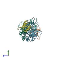
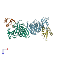
2bh1

X-ray diffraction
2.4Å resolution

X-ray structure of the general secretion pathway complex of the N- terminal domain of EpsE and the cytosolic domain of EpsL of Vibrio cholerae

Released:

Function and Biology Details

Reaction catalysed:
ATP + H(2)O + cellular protein(Side 1) = ADP + phosphate + cellular protein(Side 2)
Biochemical function:
  • not assigned

Structure analysis Details

Assembly composition:
hetero dimer (preferred)
PDBe Complex ID:
PDB-CPX-153351 (preferred)
Entry contents:
2 distinct polypeptide molecules
Macromolecules (2 distinct):
Type II secretion system protein L Chains: A, B
Molecule details ›
Chains: A, B
Length: 250 amino acids
Theoretical weight: 27.98 KDa
Source organism: Vibrio cholerae
Expression system: Escherichia coli BL21(DE3)
UniProt:
  • Canonical: P45782 (Residues: 5-246; Coverage: 60%)
Gene names: VC_2725, epsL
Sequence domains: Type II secretion system (T2SS), protein L
Structure domains:
Type II secretion system ATPase E Chains: X, Y
Molecule details ›
Chains: X, Y
Length: 96 amino acids
Theoretical weight: 11.19 KDa
Source organism: Vibrio cholerae
Expression system: Escherichia coli BL21(DE3)
UniProt:
  • Canonical: P37093 (Residues: 1-96; Coverage: 19%)
Gene names: VC_2732, epsE
Structure domains: Type II secretion system, protein E, N-terminal domain

Ligands and Environments

1 bound ligand:
No modified residues

Experiments and Validation Details

Entry percentile scores
X-ray source: ALS BEAMLINE 8.2.2
Spacegroup: P21
Unit cell:
a: 50.887Å b: 89.325Å c: 79.726Å
α: 90° β: 91.16° γ: 90°
R-values:
R R work R free
0.194 0.191 0.252
Expression system: Escherichia coli BL21(DE3)