2bk1

Electron Microscopy
29Å resolution

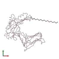
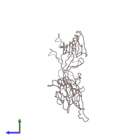
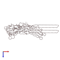
The pore structure of pneumolysin, obtained by fitting the alpha carbon trace of perfringolysin O into a cryo-EM map

Released:
Source organism: Clostridium perfringens
Related structures: EMD-1107

Function and Biology Details

Structure analysis Details

Assembly composition:
homo 38-mer (preferred)
Assembly name:
PDBe Complex ID:
PDB-CPX-142997 (preferred)
Entry contents:
1 distinct polypeptide molecule
Macromolecule:
Perfringolysin O Chain: A
Molecule details ›
Chain: A
Length: 448 amino acids
Theoretical weight: 50.1 KDa
Source organism: Clostridium perfringens
Expression system: Escherichia coli
UniProt:
  • Canonical: P0C2E9 (Residues: 53-500; Coverage: 95%)
Gene names: CPE0163, pfo, pfoA, pfoR
Sequence domains:

Ligands and Environments

No bound ligands
No modified residues

Experiments and Validation Details

Entry percentile scores
Resolution: 29Å
Relevant EMDB volumes: EMD-1107
Expression system: Escherichia coli