


Function and Biology Details
Reactions catalysed:
ATP + cytidine = ADP + CMP
ATP + inosine = ADP + IMP
Biochemical function:
Biological process:
Cellular component:
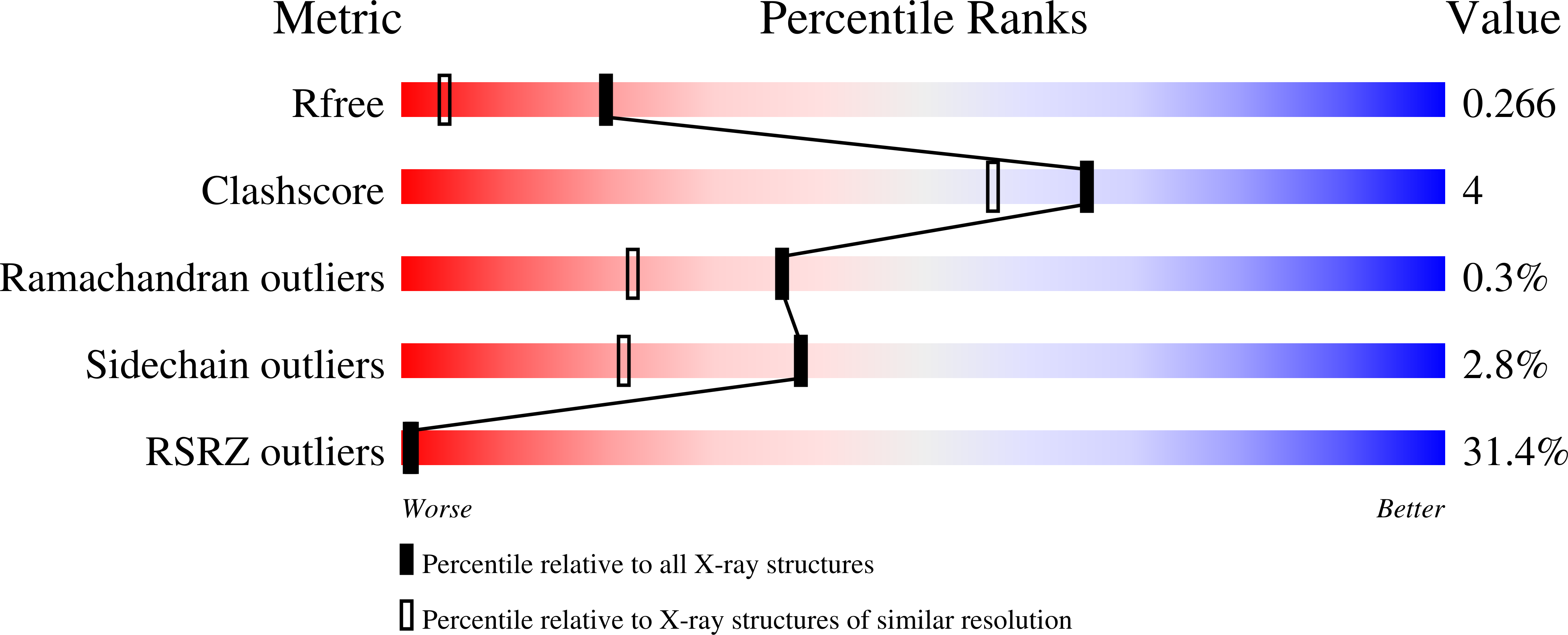
Structure analysis Details
Assembly composition:
homo dimer (preferred)
Assembly name:
Nucleoside kinase (preferred)
PDBe Complex ID:
PDB-CPX-176489 (preferred)
Entry contents:
1 distinct polypeptide molecule
Macromolecule:
Nucleoside kinase
Molecule details ›
Chain: A
Length: 302 amino acids
Theoretical weight: 33.96 KDa
Source organism: Methanocaldococcus jannaschii
Expression system: Escherichia coli BL21(DE3)
UniProt:
Sequence domains: pfkB family carbohydrate kinase
Structure domains: UDP-N-acetylmuramoyl-L-alanine:D-glutamate ligase
Length: 302 amino acids
Theoretical weight: 33.96 KDa
Source organism: Methanocaldococcus jannaschii
Expression system: Escherichia coli BL21(DE3)
UniProt:
- Canonical:
 Q57849 (Residues: 1-302; Coverage: 100%)
Sequence domains: pfkB family carbohydrate kinase
Structure domains: UDP-N-acetylmuramoyl-L-alanine:D-glutamate ligase

