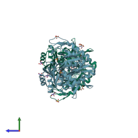
2czg

X-ray diffraction
2.35Å resolution

Crystal structure of Probable phosphoribosylglycinamide formyl transferase (PH0318) from Pyrococcus horikoshii OT3

Released:
Source organism: Pyrococcus horikoshii OT3
Entry authors: Yoshikawa S, Arai R, Kamo-Uchikubo T, Shirouzu M, Yokoyama S, RIKEN Structural Genomics/Proteomics Initiative (RSGI)

Function and Biology Details

Reaction catalysed:
ATP + formate + N(1)-(5-phospho-beta-D-ribosyl)glycinamide = ADP + phosphate + N(2)-formyl-N(1)-(5-phospho-beta-D-ribosyl)glycinamide
Biochemical function:
Cellular component:

Structure analysis Details

Assembly composition:
homo dimer (preferred)
PDBe Complex ID:
PDB-CPX-129665 (preferred)
Entry contents:
1 distinct polypeptide molecule
Macromolecule:
Formate-dependent phosphoribosylglycinamide formyltransferase Chains: A, B
Molecule details ›
Chains: A, B
Length: 433 amino acids
Theoretical weight: 48.77 KDa
Source organism: Pyrococcus horikoshii OT3
Expression system: Escherichia coli
UniProt:
  • Canonical: O58056 (Residues: 1-430; Coverage: 100%)
Gene names: PH0318, purT
Sequence domains:
Structure domains:

Ligands and Environments

2 bound ligands:
No modified residues

Experiments and Validation Details

Entry percentile scores
X-ray source: SPRING-8 BEAMLINE BL26B1
Spacegroup: C2221
Unit cell:
a: 105.465Å b: 219.285Å c: 120.753Å
α: 90° β: 90° γ: 90°
R-values:
R R work R free
0.194 0.194 0.228
Expression system: Escherichia coli