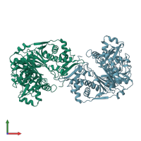
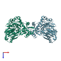
2dpn

X-ray diffraction
2.8Å resolution

Crystal Structure of the glycerol kinase from Thermus thermophilus HB8

Released:
Source organism: Thermus thermophilus
Primary publication:
Nucleant-mediated protein crystallization with the application of microporous synthetic zeolites.
Acta Crystallogr D Biol Crystallogr 64 686-95 (2008)
PMID: 18560157

Function and Biology Details

Structure analysis Details

Assembly composition:
homo dimer (preferred)
Assembly name:
PDBe Complex ID:
PDB-CPX-176141 (preferred)
Entry contents:
1 distinct polypeptide molecule
Macromolecule:
Glycerol kinase Chains: A, B
Molecule details ›
Chains: A, B
Length: 495 amino acids
Theoretical weight: 53.41 KDa
Source organism: Thermus thermophilus
Expression system: Escherichia coli BL21(DE3)
UniProt:
  • Canonical: Q53W24 (Residues: 1-495; Coverage: 100%)
Gene names: TTHB142, glpK
Sequence domains:
Structure domains: Nucleotidyltransferase; domain 5

Ligands and Environments

No bound ligands
No modified residues

Experiments and Validation Details

Entry percentile scores
X-ray source: SPRING-8 BEAMLINE BL26B1
Spacegroup: P212121
Unit cell:
a: 67.991Å b: 90.506Å c: 182.985Å
α: 90° β: 90° γ: 90°
R-values:
R R work R free
0.216 0.213 0.266
Expression system: Escherichia coli BL21(DE3)