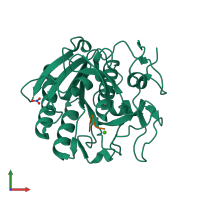
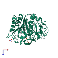
2duj

X-ray diffraction
1.67Å resolution

Crystal structure of the complex formed between proteinase K and a synthetic peptide Leu-Leu-Phe-Asn-Asp at 1.67 A resolution

Released:
Source organism: Parengyodontium album
Entry authors: Singh AK, Singh N, Somvanshi RK, Gupta D, Sharma S, Singh TP

Function and Biology Details

Reaction catalysed:
Hydrolysis of keratin, and of other proteins with subtilisin-like specificity. Hydrolyzes peptide amides.
Biochemical function:
Biological process:
Cellular component:
  • not assigned

Structure analysis Details

Assembly composition:
hetero dimer (preferred)
PDBe Complex ID:
PDB-CPX-139283 (preferred)
Entry contents:
2 distinct polypeptide molecules
Macromolecules (2 distinct):
Proteinase K Chain: A
Molecule details ›
Chain: A
Length: 279 amino acids
Theoretical weight: 28.96 KDa
Source organism: Parengyodontium album
UniProt:
  • Canonical: P06873 (Residues: 106-384; Coverage: 76%)
Gene name: PROK
Sequence domains: Subtilase family
Structure domains: Peptidase S8/S53 domain
LLFND Chain: P
Molecule details ›
Chain: P
Length: 5 amino acids
Theoretical weight: 621 Da
Source organism: Parengyodontium album
Expression system: Not provided

Ligands and Environments

2 bound ligands:
No modified residues

Experiments and Validation Details

Entry percentile scores
X-ray source: RIGAKU RU300
Spacegroup: P43212
Unit cell:
a: 68.249Å b: 68.249Å c: 108.241Å
α: 90° β: 90° γ: 90°
R-values:
R R work R free
0.17 0.163 0.192
Expression system: Not provided