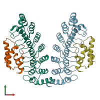
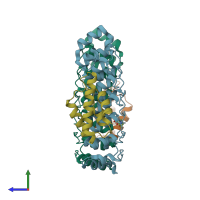
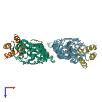
2dwz

X-ray diffraction
2.4Å resolution

Structure of the Oncoprotein Gankyrin in Complex with S6 ATPase of the 26S Proteasome

Released:
Source organisms:
Entry authors: Padmanabhan B, Yokoyama S, RIKEN Structural Genomics/Proteomics Initiative (RSGI)

Function and Biology Details

Structure analysis Details

Assembly composition:
hetero dimer (preferred)
Assembly name:
PDBe Complex ID:
PDB-CPX-179024 (preferred)
Entry contents:
2 distinct polypeptide molecules
Macromolecules (2 distinct):
26S proteasome non-ATPase regulatory subunit 10 Chains: A, C
Molecule details ›
Chains: A, C
Length: 231 amino acids
Theoretical weight: 25.09 KDa
Source organism: Mus musculus
Expression system: Escherichia coli
UniProt:
  • Canonical: Q9Z2X2 (Residues: 1-231; Coverage: 100%)
Gene name: Psmd10
Sequence domains: Ankyrin repeats (3 copies)
Structure domains: Ankyrin repeat-containing domain
26S proteasome regulatory subunit 6B Chains: B, D
Molecule details ›
Chains: B, D
Length: 83 amino acids
Theoretical weight: 9.79 KDa
Source organism: Rattus norvegicus
Expression system: Escherichia coli
UniProt:
  • Canonical: Q63570 (Residues: 337-418; Coverage: 20%)
Gene names: Psmc4, Tbp7
Sequence domains: AAA+ lid domain
Structure domains: Helicase, Ruva Protein; domain 3

Ligands and Environments

1 bound ligand:
No modified residues

Experiments and Validation Details

Entry percentile scores
X-ray source: PHOTON FACTORY BEAMLINE BL-6A
Spacegroup: P212121
Unit cell:
a: 41.991Å b: 92.319Å c: 167.191Å
α: 90° β: 90° γ: 90°
R-values:
R R work R free
0.194 0.194 0.268
Expression system: Escherichia coli