


Function and Biology Details
Reaction catalysed:
ATP + biotin + [biotin carboxyl-carrier protein]-L-lysine = AMP + diphosphate + [biotin carboxyl-carrier protein]-N(6)-biotinyl-L-lysine
Biochemical function:
Biological process:
Cellular component:
Sequence domains:
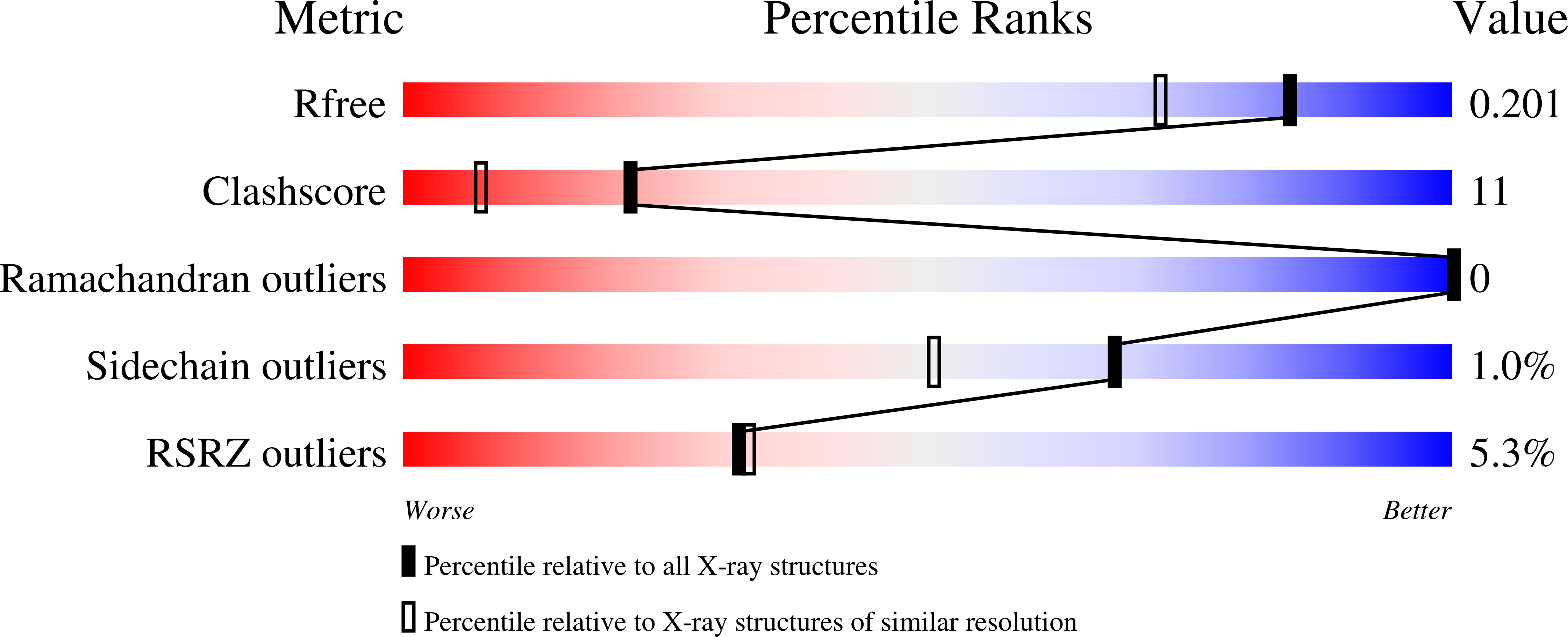
Structure analysis Details
Assembly composition:
homo dimer (preferred)
Assembly name:
BPL/LPL catalytic domain-containing protein (preferred)
PDBe Complex ID:
PDB-CPX-129627 (preferred)
Entry contents:
1 distinct polypeptide molecule
Macromolecule:
BPL/LPL catalytic domain-containing protein
Molecule details ›
Chains: A, B
Length: 235 amino acids
Theoretical weight: 26.06 KDa
Source organism: Pyrococcus horikoshii
Expression system: Escherichia coli
UniProt:
Sequence domains:
Structure domains:
Length: 235 amino acids
Theoretical weight: 26.06 KDa
Source organism: Pyrococcus horikoshii
Expression system: Escherichia coli
UniProt:
- Canonical:
 O57883 (Residues: 1-235; Coverage: 100%)
Sequence domains:
Structure domains:


