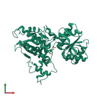
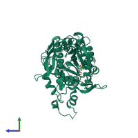
2e4z

X-ray diffraction
3.3Å resolution

Crystal structure of the ligand-binding region of the group III metabotropic glutamate receptor

Released:
Source organism: Rattus norvegicus
Primary publication:
Structures of the extracellular regions of the group II/III metabotropic glutamate receptors.
Proc Natl Acad Sci U S A 104 3759-64 (2007)
PMID: 17360426

Function and Biology Details

Structure analysis Details

Assembly composition:
homo dimer (preferred)
PDBe Complex ID:
PDB-CPX-152901 (preferred)
Entry contents:
1 distinct polypeptide molecule
Macromolecule:
Metabotropic glutamate receptor 7 Chain: A
Molecule details ›
Chain: A
Length: 501 amino acids
Theoretical weight: 56.03 KDa
Source organism: Rattus norvegicus
Expression system: Trichoplusia ni
UniProt:
  • Canonical: P35400 (Residues: 33-521; Coverage: 55%)
Gene names: Gprc1g, Grm7, Mglur7
Sequence domains: Receptor family ligand binding region
Structure domains: Rossmann fold

Ligands and Environments

1 bound ligand:
No modified residues

Experiments and Validation Details

Entry percentile scores
X-ray source: SPRING-8 BEAMLINE BL41XU
Spacegroup: P3121
Unit cell:
a: 92.431Å b: 92.431Å c: 114.332Å
α: 90° β: 90° γ: 120°
R-values:
R R work R free
0.279 0.27 0.324
Expression system: Trichoplusia ni