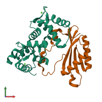
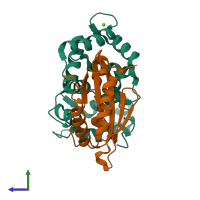
2ehb

X-ray diffraction
2.1Å resolution

The structure of the C-terminal domain of the protein kinase AtSOS2 bound to the calcium sensor AtSOS3

Released:

Function and Biology Details

Reaction catalysed:
ATP + a protein = ADP + a phosphoprotein
Biochemical function:
Biological process:
Cellular component:

Structure analysis Details

Assembly composition:
hetero dimer (preferred)
PDBe Complex ID:
PDB-CPX-131341 (preferred)
Entry contents:
2 distinct polypeptide molecules
Macromolecules (2 distinct):
Calcineurin B-like protein 4 Chain: A
Molecule details ›
Chain: A
Length: 207 amino acids
Theoretical weight: 24.05 KDa
Source organism: Arabidopsis thaliana
Expression system: Escherichia coli
UniProt:
  • Canonical: O81223 (Residues: 1-207; Coverage: 93%)
Gene names: At5g24270, CBL4, MOP9.8, SOS3
Structure domains: EF-hand
CBL-interacting serine/threonine-protein kinase 24 Chain: D
Molecule details ›
Chain: D
Length: 143 amino acids
Theoretical weight: 16.37 KDa
Source organism: Arabidopsis thaliana
Expression system: Escherichia coli
UniProt:
  • Canonical: Q9LDI3 (Residues: 304-446; Coverage: 32%)
Gene names: At5g35410, CIPK24, F6I13.1, K21B8.3, SOS2, SnRK3.11
Sequence domains: NAF domain
Structure domains: Kinase associated domain 1, KA1

Ligands and Environments

1 bound ligand:
No modified residues

Experiments and Validation Details

Entry percentile scores
X-ray source: ESRF BEAMLINE ID29
Spacegroup: P212121
Unit cell:
a: 44.137Å b: 57.387Å c: 141.896Å
α: 90° β: 90° γ: 90°
R-values:
R R work R free
0.217 0.215 0.259
Expression system: Escherichia coli