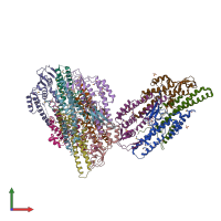
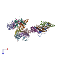
2eho

X-ray diffraction
3Å resolution

Crystal structure of human GINS complex

Released:
Source organism: Homo sapiens
Primary publication:
Crystal structure of the human GINS complex.
Genes Dev 21 1316-21 (2007)
PMID: 17545466

Function and Biology Details

Structure analysis Details

Assembly composition:
hetero tetramer (preferred)
Assembly name:
PDBe Complex ID:
PDB-CPX-172086 (preferred)
Entry contents:
4 distinct polypeptide molecules
Macromolecules (4 distinct):
DNA replication complex GINS protein SLD5 Chains: A, E, I
Molecule details ›
Chains: A, E, I
Length: 203 amino acids
Theoretical weight: 24.35 KDa
Source organism: Homo sapiens
Expression system: Escherichia coli BL21(DE3)
UniProt:
  • Canonical: Q9BRT9 (Residues: 11-213; Coverage: 91%)
Gene names: GINS4, SLD5
Sequence domains:
Structure domains:
DNA replication complex GINS protein PSF1 Chains: B, F, J
Molecule details ›
Chains: B, F, J
Length: 152 amino acids
Theoretical weight: 18.28 KDa
Source organism: Homo sapiens
Expression system: Escherichia coli BL21(DE3)
UniProt:
  • Canonical: Q14691 (Residues: 1-151; Coverage: 77%)
Gene names: GINS1, KIAA0186, PSF1
Sequence domains: GINS complex protein
Structure domains: Methane Monooxygenase Hydroxylase; Chain G, domain 1
DNA replication complex GINS protein PSF2 Chains: C, G, K
Molecule details ›
Chains: C, G, K
Length: 186 amino acids
Theoretical weight: 22.01 KDa
Source organism: Homo sapiens
Expression system: Escherichia coli BL21(DE3)
UniProt:
  • Canonical: Q9Y248 (Residues: 1-185; Coverage: 100%)
Gene names: CGI-122, DC5, GINS2, HSPC037, PSF2
Sequence domains: GINS complex protein
Structure domains:
DNA replication complex GINS protein PSF3 Chains: D, H, L
Molecule details ›
Chains: D, H, L
Length: 216 amino acids
Theoretical weight: 24.84 KDa
Source organism: Homo sapiens
Expression system: Escherichia coli BL21(DE3)
UniProt:
  • Canonical: Q9BRX5 (Residues: 1-216; Coverage: 100%)
Gene names: GINS3, PSF3
Sequence domains: GINS complex protein
Structure domains: Methane Monooxygenase Hydroxylase; Chain G, domain 1

Ligands and Environments

1 bound ligand:
1 modified residue:

Experiments and Validation Details

Entry percentile scores
X-ray source: PAL/PLS BEAMLINE 4A
Spacegroup: C2
Unit cell:
a: 164.935Å b: 176.995Å c: 126.508Å
α: 90° β: 105.91° γ: 90°
R-values:
R R work R free
0.25 0.221 0.274
Expression system: Escherichia coli BL21(DE3)