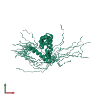
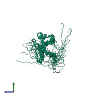
Function and Biology Details
Reaction catalysed:
(1a) a [histone H3]-N(6),N(6),N(6)-trimethyl-L-lysine(4) + 2-oxoglutarate + O(2) = a [histone H3]-N(6),N(6)-dimethyl-L-lysine(4) + succinate + formaldehyde + CO(2)
Biochemical function:
Biological process:
Cellular component:
Structure analysis Details
Assembly composition:
monomeric (preferred)
Assembly name:
Lysine-specific demethylase 5B (preferred)
PDBe Complex ID:
PDB-CPX-182408 (preferred)
Entry contents:
1 distinct polypeptide molecule
Macromolecule:
Ligands and Environments
No bound ligands
No modified residues
Experiments and Validation Details
Refinement method:
torsion angle dyanamics, simulated annealing
Expression system: Not provided


