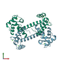
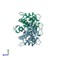
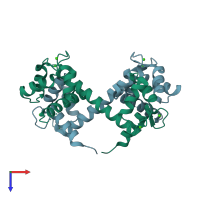
2f2p

X-ray diffraction
2.6Å resolution

Structure of calmodulin bound to a calcineurin peptide: a new way of making an old binding mode

Released:
Source organism: Bos taurus

Function and Biology Details

Reaction catalysed:
[a protein]-serine/threonine phosphate + H(2)O = [a protein]-serine/threonine + phosphate

Structure analysis Details

Assemblies composition:
homo dimer (preferred)
homo tetramer
PDBe Complex ID:
PDB-CPX-155735 (preferred)
Entry contents:
1 distinct polypeptide molecule
Macromolecule:
Calmodulin; Protein phosphatase 3 catalytic subunit alpha Chains: A, B
Molecule details ›
Chains: A, B
Length: 179 amino acids
Theoretical weight: 19.93 KDa
Source organism: Bos taurus
Expression system: Escherichia coli
UniProt:
  • Canonical: P62157 (Residues: 1-149; Coverage: 100%)
  • Canonical: P48452 (Residues: 389-413; Coverage: 5%)
Gene names: CALM, CAM, PPP3CA
Sequence domains: EF-hand domain pair
Structure domains: EF-hand

Ligands and Environments

1 bound ligand:
No modified residues

Experiments and Validation Details

Entry percentile scores
X-ray source: NSLS BEAMLINE X8C
Spacegroup: P321
Unit cell:
a: 139.549Å b: 139.549Å c: 45.592Å
α: 90° β: 90° γ: 120°
R-values:
R R work R free
0.24 0.234 0.297
Expression system: Escherichia coli