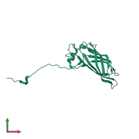
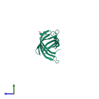
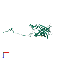
2fgs

X-ray diffraction
2.9Å resolution

Crystal structure of Campylobacter jejuni YCEI protein, structural genomics

Released:
Source organism: Campylobacter jejuni
Entry authors: Patskovsky Y, Ramagopal U, Almo SC, Burley SK, New York SGX Research Center for Structural Genomics (NYSGXRC)

Function and Biology Details

Biochemical function:
  • not assigned
Biological process:
  • not assigned
Cellular component:
  • not assigned

Structure analysis Details

Assembly composition:
homo dimer (preferred)
PDBe Complex ID:
PDB-CPX-181268 (preferred)
Entry contents:
1 distinct polypeptide molecule
Macromolecule:
Lipid/polyisoprenoid-binding YceI-like domain-containing protein Chain: A
Molecule details ›
Chain: A
Length: 179 amino acids
Theoretical weight: 20.07 KDa
Source organism: Campylobacter jejuni
Expression system: Escherichia coli BL21(DE3)
UniProt:
  • Canonical: Q79JB5 (Residues: 22-190; Coverage: 100%)
Gene names: B7A03_01670, BFD99_04285, C1418_04240, DDV78_03610, E7N58_02955, E8P16_03870, FBF07_00895, GNO00_03675, NCTC13105_01146, QE916_001052, yceI
Sequence domains: YceI-like domain
Structure domains: Lipid/polyisoprenoid-binding, YceI-like

Ligands and Environments

1 bound ligand:
No modified residues

Experiments and Validation Details

Entry percentile scores
X-ray source: RIGAKU RU200
Spacegroup: P622
Unit cell:
a: 179.535Å b: 179.535Å c: 50.577Å
α: 90° β: 90° γ: 120°
R-values:
R R work R free
0.262 0.243 0.28
Expression system: Escherichia coli BL21(DE3)