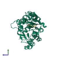
2fk8

X-ray diffraction
2Å resolution

Crystal structure of Hma (MmaA4) from Mycobacterium tuberculosis complexed with S-adenosylmethionine

Released:

Function and Biology Details

Structure analysis Details

Assembly composition:
monomeric (preferred)
PDBe Complex ID:
PDB-CPX-181263 (preferred)
Entry contents:
1 distinct polypeptide molecule
Macromolecule:
Hydroxymycolate synthase MmaA4 Chain: A
Molecule details ›
Chain: A
Length: 318 amino acids
Theoretical weight: 36.52 KDa
Source organism: Mycobacterium tuberculosis H37Rv
Expression system: Escherichia coli
UniProt:
  • Canonical: Q79FX8 (Residues: 4-301; Coverage: 99%)
Gene names: Rv0642c, hma, mma4, mmaA4
Sequence domains: Mycolic acid cyclopropane synthetase
Structure domains: Vaccinia Virus protein VP39

Ligands and Environments

1 bound ligand:
No modified residues

Experiments and Validation Details

Entry percentile scores
X-ray source: ESRF BEAMLINE ID14-4
Spacegroup: P3121
Unit cell:
a: 56.779Å b: 56.779Å c: 199.035Å
α: 90° β: 90° γ: 120°
R-values:
R R work R free
0.182 0.179 0.239
Expression system: Escherichia coli