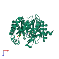
2ftp

X-ray diffraction
2.4Å resolution

Crystal Structure of hydroxymethylglutaryl-CoA lyase from Pseudomonas aeruginosa

Released:
Source organism: Pseudomonas aeruginosa PAO1
Entry authors: Xiao T, Evdokimova E, Liu Y, Kudritska M, Savchenko A, Pai EF, Edwards A, Midwest Center for Structural Genomics (MCSG)

Function and Biology Details

Reactions catalysed:
3-hydroxy-3-(4-methylpent-3-en-1-yl)glutaryl-CoA = 7-methyl-3-oxooct-6-enoyl-CoA + acetate
(S)-3-hydroxy-3-methylglutaryl-CoA = acetyl-CoA + acetoacetate
Cellular component:
  • not assigned

Structure analysis Details

Assemblies composition:
homo dimer (preferred)
homo hexamer
PDBe Complex ID:
PDB-CPX-191288 (preferred)
Entry contents:
1 distinct polypeptide molecule
Macromolecule:
3-hydroxy-3-isohexenylglutaryl-CoA/hydroxy-methylglutaryl-CoA lyase Chain: A
Molecule details ›
Chain: A
Length: 302 amino acids
Theoretical weight: 32.07 KDa
Source organism: Pseudomonas aeruginosa PAO1
Expression system: Escherichia coli BL21(DE3)
UniProt:
  • Canonical: Q9I2A0 (Residues: 1-300; Coverage: 100%)
Gene names: PA2011, liuE
Sequence domains: HMGL-like
Structure domains: Aldolase class I

Ligands and Environments

2 bound ligands:
No modified residues

Experiments and Validation Details

Entry percentile scores
X-ray source: RIGAKU FR-C
Spacegroup: I432
Unit cell:
a: 189.012Å b: 189.012Å c: 189.012Å
α: 90° β: 90° γ: 90°
R-values:
R R work R free
0.188 0.187 0.213
Expression system: Escherichia coli BL21(DE3)