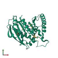
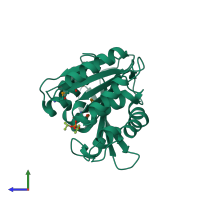
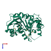
2gzs

X-ray diffraction
1.4Å resolution

Enterobactin Hydolase IroE Complex with DFP

Released:
Source organism: Escherichia coli
Primary publication:
Structural characterization of enterobactin hydrolase IroE.
Biochemistry 45 10184-90 (2006)
PMID: 16922493

Function and Biology Details

Biological process:
  • not assigned
Cellular component:
  • not assigned
Structure domain:

Structure analysis Details

Assembly composition:
monomeric (preferred)
PDBe Complex ID:
PDB-CPX-179832 (preferred)
Entry contents:
1 distinct polypeptide molecule
Macromolecule:
Catecholate siderophore esterase IroE Chain: A
Molecule details ›
Chain: A
Length: 278 amino acids
Theoretical weight: 31.01 KDa
Source organism: Escherichia coli
Expression system: Escherichia coli
UniProt:
  • Canonical: Q6KD95 (Residues: 41-318; Coverage: 97%)
Gene names: FPI65_12510, FVB16_05500, iroE
Sequence domains: Putative esterase
Structure domains: alpha/beta hydrolase

Ligands and Environments

1 bound ligand:
1 modified residue:

Experiments and Validation Details

Entry percentile scores
X-ray source: ALS BEAMLINE 8.2.1
Spacegroup: P1
Unit cell:
a: 36.295Å b: 38.361Å c: 44.631Å
α: 113.1° β: 93.99° γ: 98.08°
R-values:
R R work R free
0.174 0.174 0.186
Expression system: Escherichia coli