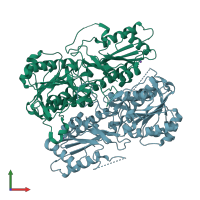
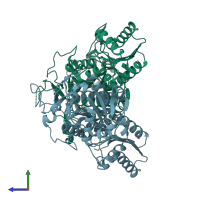
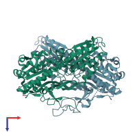
2hig

X-ray diffraction
2.4Å resolution

Crystal Structure of Phosphofructokinase apoenzyme from Trypanosoma brucei.

Released:

Function and Biology Details

Reaction catalysed:
ATP + beta-D-fructose 6-phosphate = ADP + beta-D-fructose 1,6-bisphosphate
Biochemical function:
Cellular component:

Structure analysis Details

Assembly composition:
homo tetramer (preferred)
PDBe Complex ID:
PDB-CPX-127542 (preferred)
Entry contents:
1 distinct polypeptide molecule
Macromolecule:
ATP-dependent 6-phosphofructokinase Chains: A, B
Molecule details ›
Chains: A, B
Length: 487 amino acids
Theoretical weight: 53.59 KDa
Source organism: Trypanosoma brucei
Expression system: Escherichia coli
UniProt:
  • Canonical: O15648 (Residues: 1-487; Coverage: 100%)
Gene name: pfk
Sequence domains: Phosphofructokinase
Structure domains:

Ligands and Environments

1 bound ligand:
No modified residues

Experiments and Validation Details

Entry percentile scores
X-ray source: SRS BEAMLINE PX14.1
Spacegroup: P21212
Unit cell:
a: 118.765Å b: 92.497Å c: 79.534Å
α: 90° β: 90° γ: 90°
R-values:
R R work R free
0.201 0.201 0.238
Expression system: Escherichia coli