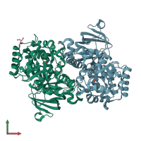
2i9u

X-ray diffraction
2.05Å resolution

Crystal Structure of Guanine Deaminase from C. acetobutylicum with bound guanine in the active site

Released:
Entry authors: Kumaran D, Burley SK, Swaminathan S, New York SGX Research Center for Structural Genomics (NYSGXRC)

Function and Biology Details

Reaction catalysed:
Guanine + H(2)O = xanthine + NH(3)
Biochemical function:
Biological process:
Cellular component:

Structure analysis Details

Assembly composition:
homo dimer (preferred)
Assembly name:
PDBe Complex ID:
PDB-CPX-188938 (preferred)
Entry contents:
1 distinct polypeptide molecule
Macromolecule:
guanine deaminase Chains: A, B
Molecule details ›
Chains: A, B
Length: 439 amino acids
Theoretical weight: 49.94 KDa
Source organism: Clostridium acetobutylicum ATCC 824
Expression system: Escherichia coli
UniProt:
  • Canonical: Q97MB6 (Residues: 2-428; Coverage: 100%)
Gene name: CA_C0282
Sequence domains: Amidohydrolase family
Structure domains:

Ligands and Environments

3 bound ligands:
No modified residues

Experiments and Validation Details

Entry percentile scores
X-ray source: NSLS BEAMLINE X29A
Spacegroup: P212121
Unit cell:
a: 87.385Å b: 88.994Å c: 139.596Å
α: 90° β: 90° γ: 90°
R-values:
R R work R free
0.194 0.194 0.224
Expression system: Escherichia coli