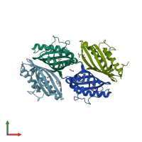
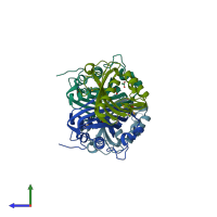
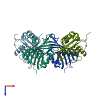
2imj

X-ray diffraction
1.5Å resolution

X-ray Crystal Structure of Protein PFL_3262 from Pseudomonas fluorescens. Northeast Structural Genomics Consortium Target PlR14.

Released:
Source organism: Pseudomonas protegens Pf-5
Entry authors: Zhou W, Forouhar F, Seetharaman J, Chen CX, Fang Y, Cunningham K, Ma L-C, Xiao R, Liu J, Baran MC, Acton TB, Montelione GT, Hunt JF, Tong L, Northeast Structural Genomics Consortium (NESG)

Function and Biology Details

Biochemical function:
  • not assigned
Biological process:
  • not assigned
Cellular component:
  • not assigned

Structure analysis Details

Assembly composition:
homo dimer (preferred)
PDBe Complex ID:
PDB-CPX-175613 (preferred)
Entry contents:
1 distinct polypeptide molecule
Macromolecule:
50S ribosomal protein L21 Chains: A, B, C, D
Molecule details ›
Chains: A, B, C, D
Length: 166 amino acids
Theoretical weight: 20.11 KDa
Source organism: Pseudomonas protegens Pf-5
Expression system: Escherichia coli
UniProt:
  • Canonical: Q4KBL6 (Residues: 1-158; Coverage: 99%)
Gene name: PFL_3262
Sequence domains: Protein of unknown function (DUF1348)
Structure domains: Nuclear Transport Factor 2; Chain: A,

Ligands and Environments

2 bound ligands:
1 modified residue:

Experiments and Validation Details

Entry percentile scores
X-ray source: NSLS BEAMLINE X4A
Spacegroup: P21
Unit cell:
a: 51.858Å b: 88.76Å c: 74.704Å
α: 90° β: 103.26° γ: 90°
R-values:
R R work R free
0.171 0.169 0.193
Expression system: Escherichia coli