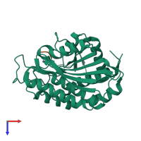
2iq6

X-ray diffraction
2Å resolution

Crystal Structure of the Aminopeptidase from Vibrio proteolyticus in Complexation with Leucyl-leucyl-leucine.

Released:

Function and Biology Details

Reaction catalysed:
Release of an N-terminal amino acid, preferentially leucine, but not glutamic or aspartic acids.
Biochemical function:
Biological process:
Cellular component:
  • not assigned

Structure analysis Details

Assembly composition:
hetero dimer (preferred)
PDBe Complex ID:
PDB-CPX-162762 (preferred)
Entry contents:
2 distinct polypeptide molecules
Macromolecules (2 distinct):
Bacterial leucyl aminopeptidase Chain: A
Molecule details ›
Chain: A
Length: 291 amino acids
Theoretical weight: 31.43 KDa
Source organism: Vibrio proteolyticus
UniProt:
  • Canonical: Q01693 (Residues: 107-397; Coverage: 60%)
Sequence domains: Peptidase family M28
Structure domains: Zn peptidases
Peptide, (Leucyl-leucyl-leucine) Chain: B
Molecule details ›
Chain: B
Length: 3 amino acids
Theoretical weight: 357 Da
Source organism: synthetic construct
Expression system: Not provided

Ligands and Environments

1 bound ligand:
No modified residues

Experiments and Validation Details

Entry percentile scores
X-ray source: RIGAKU
Spacegroup: P6122
Unit cell:
a: 108.4Å b: 108.4Å c: 96.8Å
α: 90° β: 90° γ: 120°
R-values:
R R work R free
0.206 0.206 0.247
Expression system: Not provided