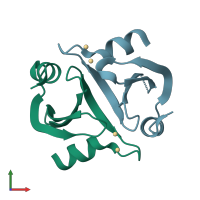
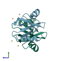
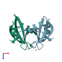
2iy2

X-ray diffraction
1.9Å resolution

Crystal structure of the N-terminal dimer domain of E.coli DsbG

Released:
Source organism: Escherichia coli
Primary publication:
Structures of the dimerization domains of the Escherichia coli disulfide-bond isomerase enzymes DsbC and DsbG.
Acta Crystallogr D Biol Crystallogr 63 465-71 (2007)
PMID: 17372350

Function and Biology Details

Biochemical function:
  • not assigned
Biological process:
  • not assigned
Cellular component:

Structure analysis Details

Assembly composition:
homo dimer (preferred)
PDBe Complex ID:
PDB-CPX-160127 (preferred)
Entry contents:
1 distinct polypeptide molecule
Macromolecule:
Thiol:disulfide interchange protein DsbG Chains: A, B
Molecule details ›
Chains: A, B
Length: 72 amino acids
Theoretical weight: 7.93 KDa
Source organism: Escherichia coli
UniProt:
  • Canonical: P77202 (Residues: 19-89; Coverage: 31%)
Gene names: JW0597, b0604, dsbG, ybdP
Structure domains: Disulphide bond isomerase, DsbC/G, N-terminal

Ligands and Environments

1 bound ligand:
No modified residues

Experiments and Validation Details

Entry percentile scores
X-ray source: SLS BEAMLINE X06SA
Spacegroup: P41
Unit cell:
a: 56.842Å b: 56.842Å c: 42.049Å
α: 90° β: 90° γ: 90°
R-values:
R R work R free
0.198 0.196 0.236