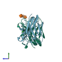
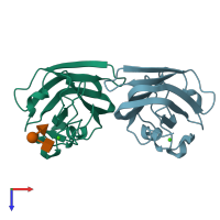
2j1v

X-ray diffraction
1.45Å resolution

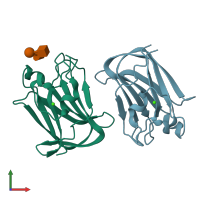
Structure of a Streptococcus pneumoniae fucose binding module in complex with the blood group H-trisaccharide

Released:
Primary publication:
Blood group antigen recognition by a Streptococcus pneumoniae virulence factor.
J Biol Chem 281 35263-71 (2006)
PMID: 16987809

Function and Biology Details

Biochemical function:
  • not assigned
Biological process:
  • not assigned
Cellular component:
  • not assigned

Structure analysis Details

Assembly composition:
monomeric (preferred)
PDBe Complex ID:
PDB-CPX-101490 (preferred)
Entry contents:
1 distinct polypeptide molecule
Macromolecules (2 distinct):
F5/8 type C domain-containing protein Chains: A, B
Molecule details ›
Chains: A, B
Length: 151 amino acids
Theoretical weight: 16.85 KDa
Source organism: Streptococcus pneumoniae TIGR4
Expression system: Escherichia coli
UniProt:
  • Canonical: A0A0H2US34 (Residues: 601-745; Coverage: 14%)
Gene name: SP_2159
Sequence domains: F5/8 type C domain
Structure domains: Galactose-binding domain-like

Ligands and Environments

Carbohydrate polymer : NEW Components: NAG, GAL, FUC
1 bound ligand:
No modified residues

Experiments and Validation Details

Entry percentile scores
Spacegroup: P212121
Unit cell:
a: 43.827Å b: 60.465Å c: 99.498Å
α: 90° β: 90° γ: 90°
R-values:
R R work R free
0.154 0.152 0.208
Expression system: Escherichia coli