Function and Biology Details
Reactions catalysed:
GTP + a 5'-diphospho-[mRNA] = diphosphate + a 5'-(5'-triphosphoguanosine)-[mRNA]
Thiol-dependent hydrolysis of ester, thioester, amide, peptide and isopeptide bonds formed by the C-terminal Gly of ubiquitin (a 76-residue protein attached to proteins as an intracellular targeting signal).
TSAVLQ-|-SGFRK-NH(2) and SGVTFQ-|-GKFKK the two peptides corresponding to the two self-cleavage sites of the SARS 3C-like proteinase are the two most reactive peptide substrates. The enzyme exhibits a strong preference for substrates containing Gln at P1 position and Leu at P2 position.
Biochemical function:
Biological process:
- not assigned
Cellular component:
- not assigned
Structure analysis Details
Assembly composition:
monomeric (preferred)
Assembly name:
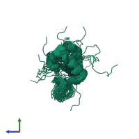
Papain-like protease nsp3 (preferred)
PDBe Complex ID:
PDB-CPX-143085 (preferred)
Entry contents:
1 distinct polypeptide molecule
Macromolecule:
Ligands and Environments
No bound ligands
No modified residues
Experiments and Validation Details
Chemical shift assignment:
83%
Refinement method:
simulated annealing
Expression system: Escherichia coli


