


Function and Biology Details
Biochemical function:
Biological process:
Cellular component:
Sequence domains:
Structure domain:
Structure analysis Details
Assembly composition:
homo 180-mer (preferred)
Assembly name:
Capsid protein (preferred)
PDBe Complex ID:
PDB-CPX-137135 (preferred)
Entry contents:
1 distinct polypeptide molecule
Macromolecule:
Capsid protein
Molecule details ›
Chains: A, B, C
Length: 129 amino acids
Theoretical weight: 13.74 KDa
Source organism: Escherichia phage MS2
Expression system: Not provided
UniProt:
Structure domains: MS2 Viral Coat Protein
Length: 129 amino acids
Theoretical weight: 13.74 KDa
Source organism: Escherichia phage MS2
Expression system: Not provided
UniProt:
- Canonical:
 P03612 (Residues: 2-130; Coverage: 99%)
Structure domains: MS2 Viral Coat Protein
Ligands and Environments
No bound ligands
No modified residues
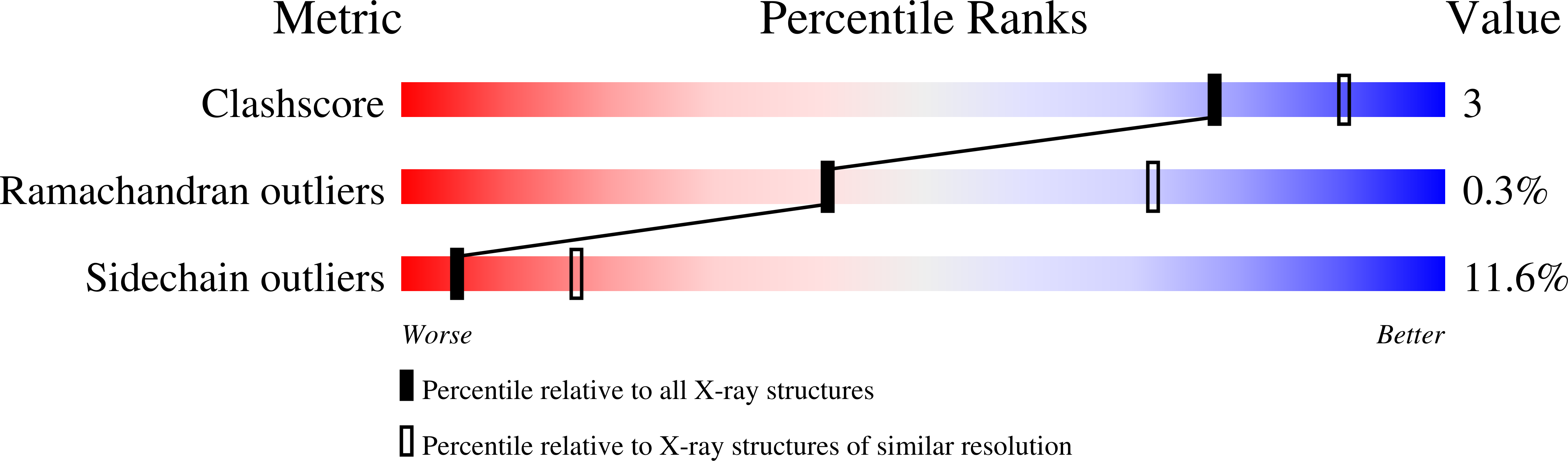
Experiments and Validation Details

Spacegroup:
R32
Expression system: Not provided