


Function and Biology Details
Reaction catalysed:
Cleaves type-1 transmembrane domains using a catalytic dyad composed of serine and histidine that are contributed by different transmembrane domains.
Biochemical function:
Biological process:
Cellular component:
Sequence domains:
Structure domain:
Structure analysis Details
Assembly composition:
homo dimer (preferred)
Assembly name:
Rhomboid protease GlpG (preferred)
PDBe Complex ID:
PDB-CPX-140641 (preferred)
Entry contents:
1 distinct polypeptide molecule
Macromolecule:
Rhomboid protease GlpG
Molecule details ›
Chains: A, B
Length: 182 amino acids
Theoretical weight: 20.53 KDa
Source organism: Escherichia coli
Expression system: Escherichia coli
UniProt:
Sequence domains: Rhomboid domain
Structure domains: Rhomboid-like
Length: 182 amino acids
Theoretical weight: 20.53 KDa
Source organism: Escherichia coli
Expression system: Escherichia coli
UniProt:
- Canonical:
 P09391 (Residues: 91-272; Coverage: 66%)
Sequence domains: Rhomboid domain
Structure domains: Rhomboid-like
Ligands and Environments
No bound ligands
No modified residues
Experiments and Validation Details
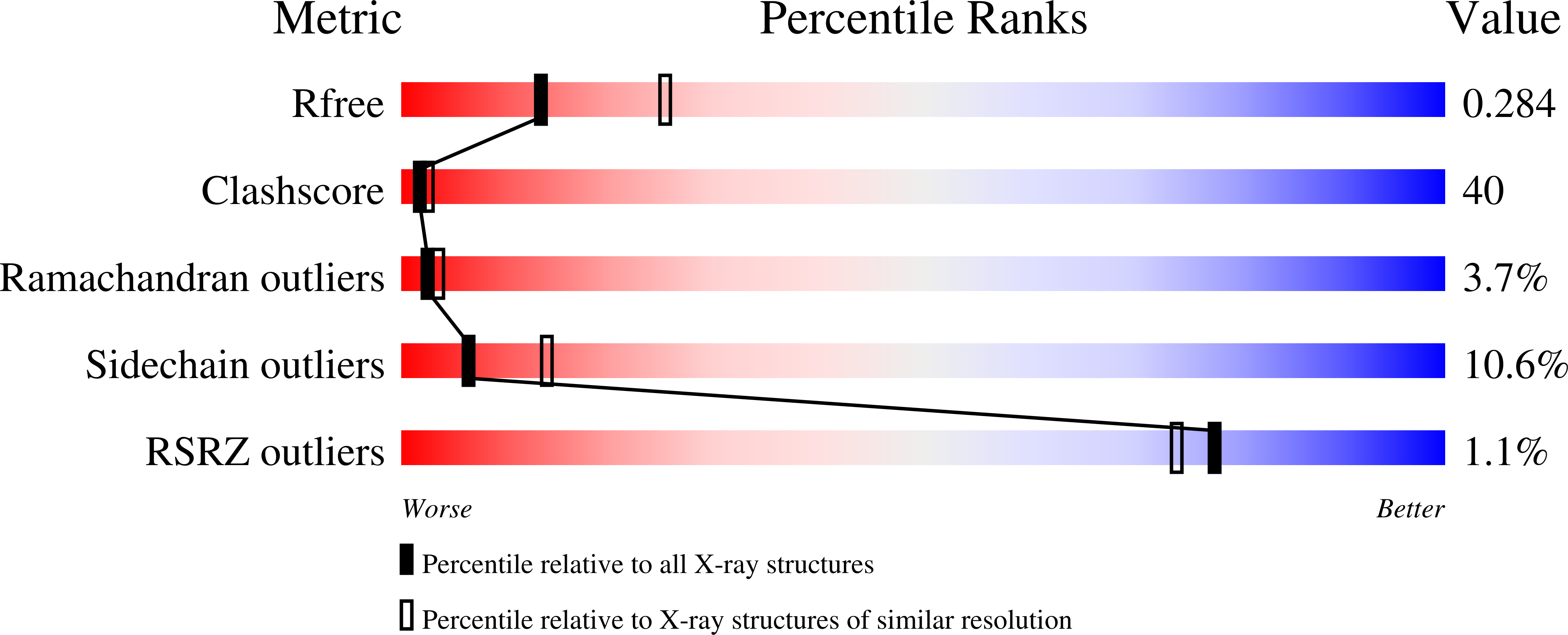
wwPDB Validation report is not available for this entry.
X-ray source:
NSLS BEAMLINE X29A
Spacegroup:
P31
Expression system: Escherichia coli
