


Function and Biology Details
Biochemical function:
Biological process:
Cellular component:
Sequence domains:
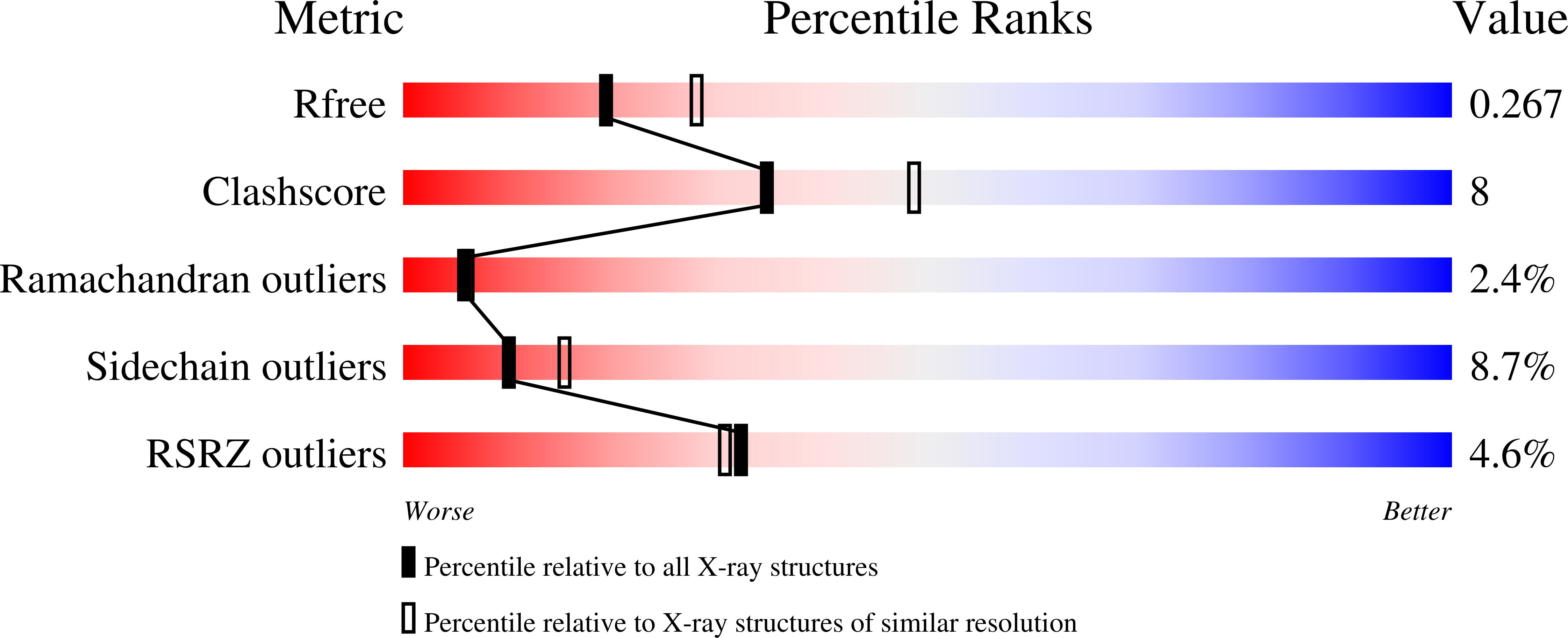
Structure analysis Details
Assemblies composition:
Assembly name:
Calcium-binding protein 1 (preferred)
PDBe Complex ID:
PDB-CPX-153663 (preferred)
Entry contents:
1 distinct polypeptide molecule
Macromolecule:
Calcium-binding protein 1
Molecule details ›
Chains: A, B
Length: 134 amino acids
Theoretical weight: 14.97 KDa
Source organism: Entamoeba histolytica HM-1:IMSS
Expression system: Escherichia coli BL21
UniProt:
Sequence domains: EF-hand domain pair
Structure domains: EF-hand
Length: 134 amino acids
Theoretical weight: 14.97 KDa
Source organism: Entamoeba histolytica HM-1:IMSS
Expression system: Escherichia coli BL21
UniProt:
- Canonical:
 P38505 (Residues: 1-134; Coverage: 100%)
Sequence domains: EF-hand domain pair
Structure domains: EF-hand


