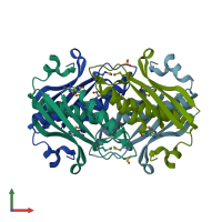
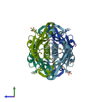
2ogf

X-ray diffraction
1.89Å resolution

Crystal structure of protein MJ0408 from Methanococcus jannaschii, Pfam DUF372

Released:
Entry authors: Ramagopal UA, Toro R, Sauder JM, Burley SK, Almo SC, New York SGX Research Center for Structural Genomics (NYSGXRC)

Function and Biology Details

Reaction catalysed:
7,8-dihydroneopterin = 6-hydroxymethyl-7,8-dihydropterin + glycolaldehyde
Biochemical function:
Cellular component:
  • not assigned

Structure analysis Details

Assembly composition:
homo tetramer (preferred)
PDBe Complex ID:
PDB-CPX-176490 (preferred)
Entry contents:
1 distinct polypeptide molecule
Macromolecule:
Dihydroneopterin aldolase Chains: A, B, C, D
Molecule details ›
Chains: A, B, C, D
Length: 122 amino acids
Theoretical weight: 14.41 KDa
Source organism: Methanocaldococcus jannaschii
Expression system: Escherichia coli BL21(DE3)
UniProt:
  • Canonical: Q57851 (Residues: 2-121; Coverage: 99%)
Gene names: MJ0408, mptD
Sequence domains: Dihydroneopterin aldolase
Structure domains: 7,8-dihydroneopterin aldolase (MptD)

Ligands and Environments

3 bound ligands:
1 modified residue:

Experiments and Validation Details

Entry percentile scores
X-ray source: OTHER
Spacegroup: P21
Unit cell:
a: 75.693Å b: 46.27Å c: 79.276Å
α: 90° β: 118.54° γ: 90°
R-values:
R R work R free
0.175 0.173 0.224
Expression system: Escherichia coli BL21(DE3)