2osl

X-ray diffraction
2.6Å resolution

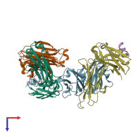
Crystal structure of Rituximab Fab in complex with an epitope peptide

Released:
Source organisms:
Primary publication:
Structural basis for recognition of CD20 by therapeutic antibody Rituximab.
J Biol Chem 282 15073-80 (2007)
PMID: 17395584

Function and Biology Details

Structure analysis Details

Assembly composition:
hetero trimer (preferred)
PDBe Complex ID:
PDB-CPX-233281 (preferred)
Entry contents:
3 distinct polypeptide molecules
Macromolecules (3 distinct):
light chain of the Rituximab Fab fragment,light chain of the Rituximab Fab fragment Chains: B, L
Molecule details ›
Chains: B, L
Length: 213 amino acids
Theoretical weight: 23.08 KDa
Source organism: Mus musculus
Expression system: synthetic construct
Structure domains: Immunoglobulins
heavy chain of the Rituximab Fab fragment,heavy chain of the Rituximab Fab fragment Chains: A, H
Molecule details ›
Chains: A, H
Length: 224 amino acids
Theoretical weight: 23.73 KDa
Source organism: Mus musculus
Expression system: synthetic construct
Structure domains: Immunoglobulins
B-lymphocyte antigen CD20 Chains: P, Q
Molecule details ›
Chains: P, Q
Length: 25 amino acids
Theoretical weight: 2.85 KDa
Source organism: Homo sapiens
Expression system: Not provided
UniProt:
  • Canonical: P11836 (Residues: 163-187; Coverage: 8%)
Gene names: CD20, MS4A1

Ligands and Environments

No bound ligands
No modified residues

Experiments and Validation Details

Entry percentile scores
X-ray source: PHOTON FACTORY BEAMLINE AR-NW12A
Spacegroup: P212121
Unit cell:
a: 96.421Å b: 98.809Å c: 107.241Å
α: 90° β: 90° γ: 90°
R-values:
R R work R free
0.239 0.239 0.296
Expression systems:
  • synthetic construct
  • Not provided