2p4n

Electron Microscopy
9Å resolution

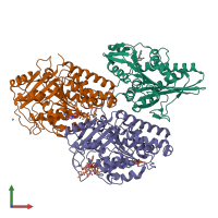
Human Monomeric Kinesin (1BG2) and Bovine Tubulin (1JFF) Docked into the 9-Angstrom Cryo-EM Map of Nucleotide-Free Kinesin Complexed to the Microtubule

Released:
Primary publication:
The beginning of kinesin's force-generating cycle visualized at 9-A resolution.
J Cell Biol 177 377-85 (2007)
PMID: 17470637
Related structures: EMD-1340

Function and Biology Details

Structure analysis Details

Assembly composition:
hetero trimer (preferred)
PDBe Complex ID:
PDB-CPX-135905 (preferred)
Entry contents:
3 distinct polypeptide molecules
Macromolecules (3 distinct):
Kinesin-1 heavy chain Chain: K
Molecule details ›
Chain: K
Length: 325 amino acids
Theoretical weight: 36.41 KDa
Source organism: Homo sapiens
UniProt:
  • Canonical: P33176 (Residues: 1-325; Coverage: 34%)
Gene names: KIF5B, KNS, KNS1
Sequence domains: Kinesin motor domain
Tubulin alpha-1A chain Chain: A
Molecule details ›
Chain: A
Length: 451 amino acids
Theoretical weight: 50.12 KDa
Source organism: Sus scrofa
UniProt:
  • Canonical: P02550 (Residues: 1-451; Coverage: 100%)
Gene name: TUBA1A
Sequence domains:
Tubulin beta-2B chain Chain: B
Molecule details ›
Chain: B
Length: 445 amino acids
Theoretical weight: 49.91 KDa
Source organism: Bos taurus
UniProt:
  • Canonical: Q6B856 (Residues: 1-445; Coverage: 100%)
Gene name: TUBB2B
Sequence domains:

Ligands and Environments

No modified residues

Experiments and Validation Details

Entry percentile scores
Resolution:
Relevant EMDB volumes: EMD-1340