


Function and Biology Details
Reaction catalysed:
ATP + adenosine = ADP + AMP
Biochemical function:
Biological process:
Cellular component:
Structure analysis Details
Assemblies composition:
Assembly name:
Adenosine kinase (preferred)
PDBe Complex ID:
PDB-CPX-161617 (preferred)
Entry contents:
1 distinct polypeptide molecule
Macromolecule:
Adenosine kinase
Molecule details ›
Chains: A, B
Length: 334 amino acids
Theoretical weight: 35.68 KDa
Source organism: Mycobacterium tuberculosis
Expression system: Escherichia coli
UniProt:
Sequence domains: pfkB family carbohydrate kinase
Structure domains: UDP-N-acetylmuramoyl-L-alanine:D-glutamate ligase
Length: 334 amino acids
Theoretical weight: 35.68 KDa
Source organism: Mycobacterium tuberculosis
Expression system: Escherichia coli
UniProt:
- Canonical:
 P9WID5 (Residues: 1-324; Coverage: 100%)
Sequence domains: pfkB family carbohydrate kinase
Structure domains: UDP-N-acetylmuramoyl-L-alanine:D-glutamate ligase
Ligands and Environments
No bound ligands
No modified residues
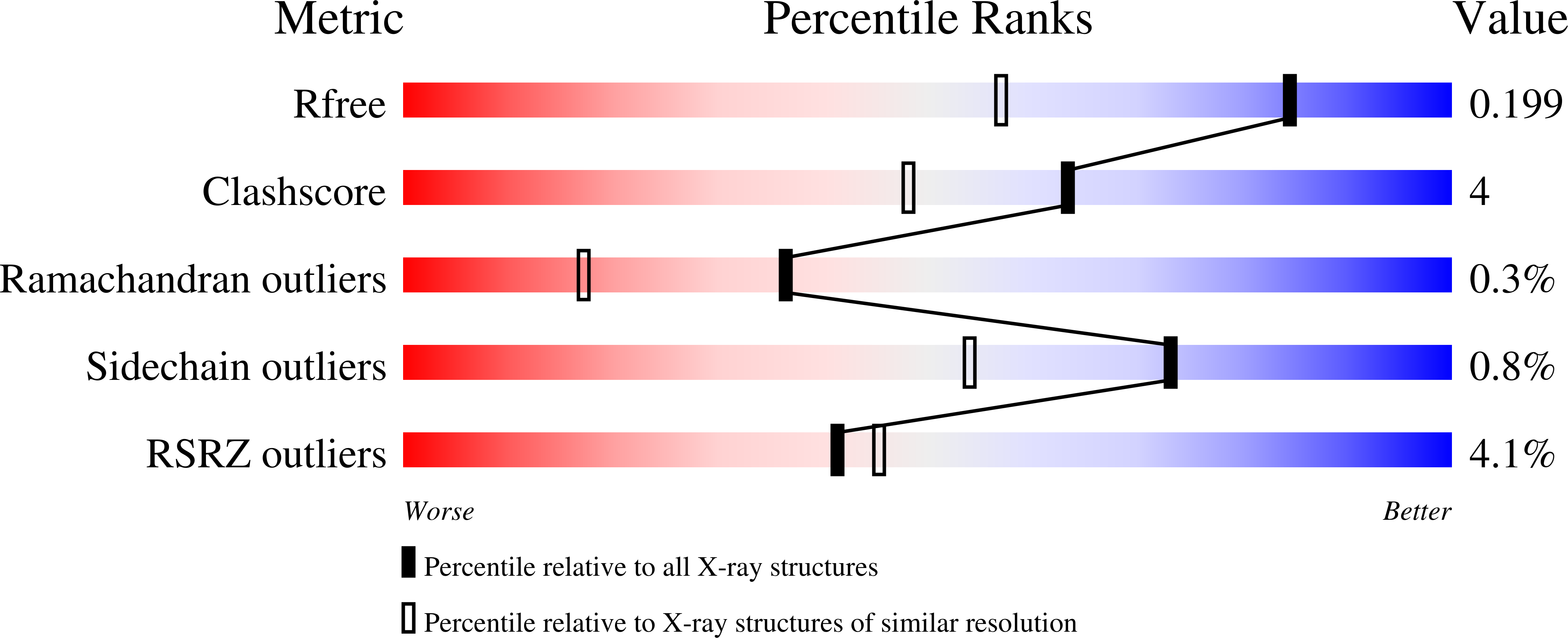
Experiments and Validation Details
wwPDB Validation report is not available for this entry.
X-ray source:
APS BEAMLINE 23-ID-B
Spacegroup:
C2
Expression system: Escherichia coli
