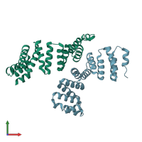
2q7f

X-ray diffraction
2.49Å resolution

Crystal structure of YrrB: a TPR protein with an unusual peptide-binding site

Released:
Source organism: Bacillus subtilis
Primary publication:
Crystal structure of YrrB: a TPR protein with an unusual peptide-binding site.
Biochem Biophys Res Commun 360 784-90 (2007)
PMID: 17624311

Function and Biology Details

Structure analysis Details

Assembly composition:
monomeric (preferred)
PDBe Complex ID:
PDB-CPX-128539 (preferred)
Entry contents:
1 distinct polypeptide molecule
Macromolecule:
TPR repeat-containing protein YrrB Chains: A, B
Molecule details ›
Chains: A, B
Length: 243 amino acids
Theoretical weight: 27.14 KDa
Source organism: Bacillus subtilis
Expression system: Escherichia coli BL21(DE3)
UniProt:
  • Canonical: O34452 (Residues: 4-202; Coverage: 97%)
Gene names: BSU27490, yrrB
Sequence domains:
Structure domains: Tetratricopeptide repeat domain

Ligands and Environments

No bound ligands
No modified residues

Experiments and Validation Details

Entry percentile scores
X-ray source: PAL/PLS BEAMLINE 4A
Spacegroup: C2
Unit cell:
a: 100.938Å b: 85.139Å c: 69.048Å
α: 90° β: 130.63° γ: 90°
R-values:
R R work R free
0.211 0.206 0.256
Expression system: Escherichia coli BL21(DE3)