2qd0

X-ray diffraction
1.81Å resolution

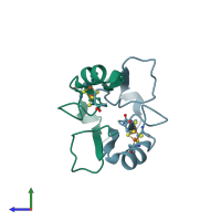
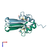
Crystal structure of mitoNEET

Released:
Source organism: Homo sapiens
Primary publication:
Crystal structure of human mitoNEET reveals distinct groups of iron sulfur proteins.
Proc Natl Acad Sci U S A 104 14640-5 (2007)
PMID: 17766439

Function and Biology Details

Reaction catalysed:
L-cysteine + 2-oxoglutarate = 2-oxo-3-sulfanylpropanoate + L-glutamate
Biochemical function:
Biological process:

Structure analysis Details

Assembly composition:
homo dimer (preferred)
PDBe Complex ID:
PDB-CPX-192731 (preferred)
Entry contents:
1 distinct polypeptide molecule
Macromolecule:
CDGSH iron-sulfur domain-containing protein 1 Chains: A, B
Molecule details ›
Chains: A, B
Length: 81 amino acids
Theoretical weight: 9.38 KDa
Source organism: Homo sapiens
Expression system: Escherichia coli BL21(DE3)
UniProt:
  • Canonical: Q9NZ45 (Residues: 32-108; Coverage: 71%)
Gene names: C10orf70, CISD1, MDS029, ZCD1
Sequence domains: Iron-binding zinc finger CDGSH type
Structure domains: CDGSH iron-sulfur domain, mitoNEET-type

Ligands and Environments

1 bound ligand:
No modified residues

Experiments and Validation Details

Entry percentile scores
X-ray source: RIGAKU MICROMAX-007, SPRING-8 BEAMLINE BL41XU
Spacegroup: P212121
Unit cell:
a: 43.838Å b: 59.139Å c: 65.971Å
α: 90° β: 90° γ: 90°
R-values:
R R work R free
0.166 0.164 0.193
Expression system: Escherichia coli BL21(DE3)