2v2f

X-ray diffraction
1.9Å resolution

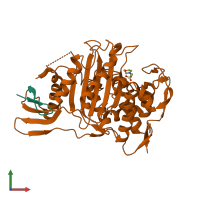
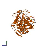
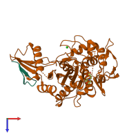
Crystal structure of PBP1a from drug-resistant strain 5204 from Streptococcus pneumoniae

Released:

Function and Biology Details

Reaction catalysed:
(GlcNAc-(1->4)-Mur2Ac(oyl-L-Ala-gamma-D-Glu-L-Lys-D-Ala-D-Ala))(n)-diphosphoundecaprenol + GlcNAc-(1->4)-Mur2Ac(oyl-L-Ala-gamma-D-Glu-L-Lys-D-Ala-D-Ala)-diphosphoundecaprenol = (GlcNAc-(1->4)-Mur2Ac(oyl-L-Ala-gamma-D-Glu-L-Lys-D-Ala-D-Ala))(n+1)-diphosphoundecaprenol + undecaprenyl diphosphate
Biochemical function:
Biological process:
  • not assigned
Cellular component:
  • not assigned

Structure analysis Details

Assembly composition:
hetero dimer (preferred)
PDBe Complex ID:
PDB-CPX-193156 (preferred)
Entry contents:
2 distinct polypeptide molecules
Macromolecules (2 distinct):
peptidoglycan glycosyltransferase Chain: A
Molecule details ›
Chain: A
Length: 24 amino acids
Theoretical weight: 2.64 KDa
Source organism: Streptococcus pneumoniae
Expression system: Escherichia coli
UniProt:
  • Canonical: Q9RET4 (Residues: 47-70; Coverage: 3%)
Gene names: pbp1A, pbp1a
peptidoglycan glycosyltransferase Chain: F
Molecule details ›
Chain: F
Length: 390 amino acids
Theoretical weight: 43.43 KDa
Source organism: Streptococcus pneumoniae
Expression system: Escherichia coli
UniProt:
  • Canonical: Q9RET4 (Residues: 264-653; Coverage: 54%)
Gene names: pbp1A, pbp1a
Sequence domains: Penicillin binding protein transpeptidase domain
Structure domains: DD-peptidase/beta-lactamase superfamily

Ligands and Environments

2 bound ligands:
No modified residues

Experiments and Validation Details

Entry percentile scores
X-ray source: ESRF BEAMLINE ID14-3
Spacegroup: C2
Unit cell:
a: 122.96Å b: 67.03Å c: 49.13Å
α: 90° β: 100.89° γ: 90°
R-values:
R R work R free
0.213 0.21 0.246
Expression system: Escherichia coli