2vf2

X-ray diffraction
2.35Å resolution

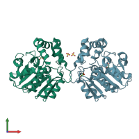
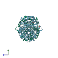
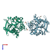
X-ray crystal structure of HsaD from Mycobacterium tuberculosis

Released:
Primary publication:
Structure of HsaD, a steroid-degrading hydrolase, from Mycobacterium tuberculosis.
Acta Crystallogr Sect F Struct Biol Cryst Commun 64 2-7 (2008)
PMID: 18097091

Function and Biology Details

Reactions catalysed:
(1E,2Z)-3-hydroxy-5,9,17-trioxo-4,5:9,10-disecoandrosta-1(10),2-dien-4-oate + H(2)O = 3-((3aS,4S,7aS)-7a-methyl-1,5-dioxo-octahydro-1H-inden-4-yl)propanoate + (2Z,4Z)-2-hydroxyhexa-2,4-dienoate
2,6-dioxo-6-phenylhexa-3-enoate + H(2)O = benzoate + 2-oxopent-4-enoate

Structure analysis Details

Assembly composition:
homo tetramer (preferred)
PDBe Complex ID:
PDB-CPX-161861 (preferred)
Entry contents:
1 distinct polypeptide molecule
Macromolecule:
4,5:9,10-diseco-3-hydroxy-5,9,17-trioxoandrosta-1(10),2-diene-4-oate hydrolase Chains: A, B
Molecule details ›
Chains: A, B
Length: 311 amino acids
Theoretical weight: 34.09 KDa
Source organism: Mycobacterium tuberculosis H37Rv
Expression system: Pseudomonas putida
UniProt:
  • Canonical: P9WNH5 (Residues: 1-291; Coverage: 100%)
Gene names: Rv3569c, bphD, hsaD
Sequence domains: alpha/beta hydrolase fold
Structure domains: alpha/beta hydrolase

Ligands and Environments

2 bound ligands:
No modified residues

Experiments and Validation Details

Entry percentile scores
X-ray source: DIAMOND BEAMLINE I04
Spacegroup: I212121
Unit cell:
a: 82.36Å b: 82.46Å c: 194.67Å
α: 90° β: 90° γ: 90°
R-values:
R R work R free
0.212 0.212 0.233
Expression system: Pseudomonas putida