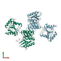
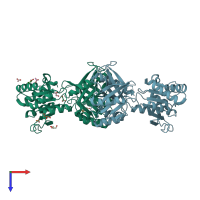
2we8

X-ray diffraction
2.3Å resolution

Crystal structure of Mycobacterium tuberculosis Rv0376c homologue from Mycobacterium smegmatis

Released:
Entry authors: Cho HJ, Kang BS

Function and Biology Details

Reaction catalysed:
Xanthine + NAD(+) + H(2)O = urate + NADH
Biochemical function:
  • not assigned
Biological process:
  • not assigned
Cellular component:
  • not assigned
Sequence domains:

Structure analysis Details

Assembly composition:
homo dimer (preferred)
Assembly name:
PDBe Complex ID:
PDB-CPX-106250 (preferred)
Entry contents:
1 distinct polypeptide molecule
Macromolecule:
Xanthine dehydrogenase Chains: A, B
Molecule details ›
Chains: A, B
Length: 386 amino acids
Theoretical weight: 40.55 KDa
Source organism: Mycolicibacterium smegmatis MC2 155
UniProt:
  • Canonical: A0QQG1 (Residues: 1-386; Coverage: 100%)
Gene name: MSMEG_0743
Sequence domains:
Structure domains: NAD(P)-binding Rossmann-like Domain

Ligands and Environments

2 bound ligands:
No modified residues

Experiments and Validation Details

Entry percentile scores
X-ray source: PAL/PLS BEAMLINE 4A
Spacegroup: P6322
Unit cell:
a: 101.208Å b: 101.208Å c: 316.669Å
α: 90° β: 90° γ: 120°
R-values:
R R work R free
0.201 0.199 0.251