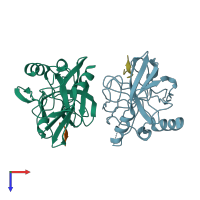
Function and Biology Details
Reactions catalysed:
Nucleoside triphosphate + RNA(n) = diphosphate + RNA(n+1)
Selective cleavage of Gln-|-Gly bond in the poliovirus polyprotein. In other picornavirus reactions Glu may be substituted for Gln, and Ser or Thr for Gly.
Autocatalytically cleaves itself from the polyprotein of the foot-and-mouth disease virus by hydrolysis of a Lys-|-Gly bond, but then cleaves host cell initiation factor eIF-4G at bonds -Gly-|-Arg- and -Lys-|-Arg-.
NTP + H(2)O = NDP + phosphate
Biochemical function:
Biological process:
Cellular component:
- not assigned
Structure analysis Details
Assembly composition:
hetero dimer (preferred)
Assembly name:
Capsid protein VP4 (preferred)
PDBe Complex ID:
PDB-CPX-136756 (preferred)
Entry contents:
2 distinct polypeptide molecules
Macromolecules (2 distinct):
Ligands and Environments
No bound ligands
No modified residues
Experiments and Validation Details
X-ray source:
ESRF BEAMLINE ID23-2
Spacegroup:
P212121
Expression systems:
- Escherichia coli BL21(DE3)
- Not provided


