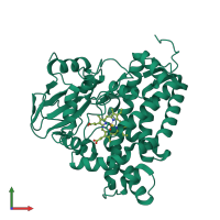
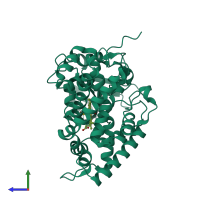
2x5l

X-ray diffraction
1.48Å resolution

X-RAY STRUCTURE OF THE SUBSTRATE-FREE MYCOBACTERIUM TUBERCULOSIS CYTOCHROME P450 CYP125, ALTERNATIVE CRYSTAL FORM

Released:
Primary publication:
Reverse type I inhibitor of Mycobacterium tuberculosis CYP125A1.
Bioorg Med Chem Lett 21 332-7 (2011)
PMID: 21109436

Function and Biology Details

Reaction catalysed:
(1a) cholest-4-en-3-one + 2 reduced ferredoxin [iron-sulfur] cluster + 2 H(+) + O(2) = (25S)-26-hydroxycholest-4-en-3-one + 2 oxidized ferredoxin [iron-sulfur] cluster + H(2)O
Biochemical function:
Cellular component:
  • not assigned

Structure analysis Details

Assembly composition:
monomeric (preferred)
PDBe Complex ID:
PDB-CPX-161999 (preferred)
Entry contents:
1 distinct polypeptide molecule
Macromolecule:
Steroid C26-monooxygenase Chain: A
Molecule details ›
Chain: A
Length: 431 amino acids
Theoretical weight: 48.33 KDa
Source organism: Mycobacterium tuberculosis H37Rv
Expression system: Escherichia coli BL21(DE3)
UniProt:
  • Canonical: P9WPP1 (Residues: 17-433; Coverage: 96%)
Gene names: MTCY03C7.11, Rv3545c, cyp125, cyp125A1
Sequence domains: Cytochrome P450
Structure domains: Cytochrome P450

Ligands and Environments

1 bound ligand:
No modified residues

Experiments and Validation Details

Entry percentile scores
X-ray source: ALS BEAMLINE 8.3.1
Spacegroup: P212121
Unit cell:
a: 54.48Å b: 74.578Å c: 102.225Å
α: 90° β: 90° γ: 90°
R-values:
R R work R free
0.153 0.151 0.202
Expression system: Escherichia coli BL21(DE3)