2xdc

X-ray diffraction
1.7Å resolution

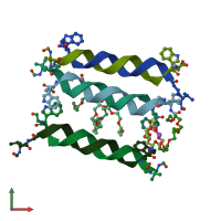
Structure of linear gramicidin D obtained using Type I crystals grown in a lipid cubic phase.

Released:
Source organism: Brevibacillus brevis
Primary publication:
Crystallizing transmembrane peptides in lipidic mesophases.
Biophys J 99 L23-5 (2010)
PMID: 20682243

Function and Biology Details

Biochemical function:
  • not assigned
Biological process:
  • not assigned
Cellular component:
  • not assigned

Structure analysis Details

Assembly composition:
homo dimer (preferred)
Assembly name:
PDBe Complex ID:
PDB-CPX-163313 (preferred)
Entry contents:
1 distinct polypeptide molecule
Macromolecule:
GRAMICIDIN A Chains: A, B, C, D, E, F
Molecule details ›
Chains: A, B, C, D, E, F
Length: 16 amino acids
Theoretical weight: 1.88 KDa
Source organism: Brevibacillus brevis

Ligands and Environments

2 bound ligands:
3 modified residues:

Experiments and Validation Details

Entry percentile scores
X-ray source: APS BEAMLINE 23-ID-B
Spacegroup: P21
Unit cell:
a: 30.5Å b: 62.56Å c: 30.53Å
α: 90° β: 99.97° γ: 90°
R-values:
R R work R free
0.181 0.179 0.214