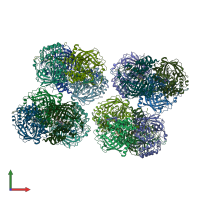
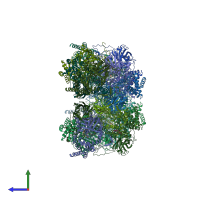
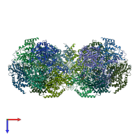
2xq1

X-ray diffraction
2.9Å resolution

Crystal structure of peroxisomal catalase from the yeast Hansenula polymorpha

Released:
Source organism: Ogataea angusta
Primary publication:
Structural features of peroxisomal catalase from the yeast Hansenula polymorpha.
Acta Crystallogr D Biol Crystallogr 67 690-8 (2011)
PMID: 21795810

Function and Biology Details

Structure analysis Details

Assembly composition:
homo tetramer (preferred)
Assembly name:
PDBe Complex ID:
PDB-CPX-151693 (preferred)
Entry contents:
1 distinct polypeptide molecule
Macromolecule:
Peroxisomal catalase Chains: A, B, C, D, E, F, G, H, I, J, K, L, M, N, O, P
Molecule details ›
Chains: A, B, C, D, E, F, G, H, I, J, K, L, M, N, O, P
Length: 509 amino acids
Theoretical weight: 58.03 KDa
Source organism: Ogataea angusta
UniProt:
  • Canonical: P30263 (Residues: 1-507; Coverage: 100%)
Gene names: PXP-9, PXP9
Sequence domains:
Structure domains: Catalase core domain

Ligands and Environments


Cofactor: Ligand HEM 15 x HEM
No bound ligands
No modified residues

Experiments and Validation Details

Entry percentile scores
X-ray source: ESRF BEAMLINE ID14-1
Spacegroup: P21
Unit cell:
a: 132.52Å b: 196.68Å c: 170.85Å
α: 90° β: 92.85° γ: 90°
R-values:
R R work R free
0.191 0.191 0.223