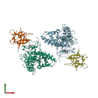
2y9w

X-ray diffraction
2.3Å resolution

Crystal structure of PPO3, a tyrosinase from Agaricus bisporus, in deoxy-form that contains additional unknown lectin-like subunit

Released:

Function and Biology Details

Reaction catalysed:
(1a) L-tyrosine + 1/2 O(2) = L-dopa
Biochemical function:
Biological process:
  • not assigned
Cellular component:
  • not assigned

Structure analysis Details

Assembly composition:
hetero dimer (preferred)
PDBe Complex ID:
PDB-CPX-111700 (preferred)
Entry contents:
2 distinct polypeptide molecules
Macromolecules (2 distinct):
Polyphenol oxidase 3 Chains: A, B
Molecule details ›
Chains: A, B
Length: 391 amino acids
Theoretical weight: 45.32 KDa
Source organism: Agaricus bisporus
UniProt:
  • Canonical: C7FF04 (Residues: 2-392; Coverage: 68%)
Gene name: PPO3
Sequence domains: Common central domain of tyrosinase
Structure domains: Di-copper center containing domain from catechol oxidase
Lectin-like fold protein Chains: C, D
Molecule details ›
Chains: C, D
Length: 150 amino acids
Theoretical weight: 16.48 KDa
Source organism: Agaricus bisporus
UniProt:
  • Canonical: G1K3P4 (Residues: 1-150; Coverage: 100%)
Structure domains: Trefoil (Acidic Fibroblast Growth Factor, subunit A)

Ligands and Environments

No modified residues

Experiments and Validation Details

Entry percentile scores
X-ray source: ESRF BEAMLINE ID14-4
Spacegroup: P21212
Unit cell:
a: 104.06Å b: 104.52Å c: 109.05Å
α: 90° β: 90° γ: 90°
R-values:
R R work R free
0.184 0.181 0.237