2z6b

X-ray diffraction
3.11Å resolution

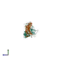
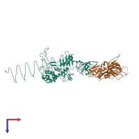
Crystal Structure Analysis of (gp27-gp5)3 conjugated with Fe(III) protoporphyrin

Released:

Function and Biology Details

Reaction catalysed:
Hydrolysis of (1->4)-beta-linkages between N-acetylmuramic acid and N-acetyl-D-glucosamine residues in a peptidoglycan and between N-acetyl-D-glucosamine residues in chitodextrins
Biochemical function:
Biological process:
Cellular component:

Structure analysis Details

Assembly composition:
hetero hexamer (preferred)
PDBe Complex ID:
PDB-CPX-147582 (preferred)
Entry contents:
2 distinct polypeptide molecules
Macromolecules (2 distinct):
Pre-baseplate central spike protein Gp5 Chain: A
Molecule details ›
Chain: A
Length: 584 amino acids
Theoretical weight: 64.33 KDa
Source organism: Tequatrovirus T4
Expression system: Escherichia coli BL21(DE3)
UniProt:
  • Canonical: P16009 (Residues: 1-575; Coverage: 100%)
Gene name: 5
Sequence domains:
Structure domains:
Baseplate central spike complex protein gp27 Chain: D
Molecule details ›
Chain: D
Length: 391 amino acids
Theoretical weight: 44.43 KDa
Source organism: Tequatrovirus T4
Expression system: Escherichia coli BL21(DE3)
UniProt:
  • Canonical: P17172 (Residues: 1-391; Coverage: 100%)
Gene name: 27
Sequence domains:
Structure domains:

Ligands and Environments

1 bound ligand:
No modified residues

Experiments and Validation Details

Entry percentile scores
X-ray source: SPRING-8 BEAMLINE BL44B2
Spacegroup: R32
Unit cell:
a: 138.536Å b: 138.536Å c: 389.121Å
α: 90° β: 90° γ: 120°
R-values:
R R work R free
0.243 0.243 0.315
Expression system: Escherichia coli BL21(DE3)