2z9o

X-ray diffraction
3.14Å resolution

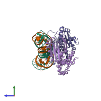
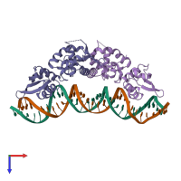
Crystal structure of the dimeric form of RepE in complex with the repE operator DNA

Released:
Source organism: Escherichia coli K-12
Primary publication:
Structural basis for regulation of bifunctional roles in replication initiator protein.
Proc Natl Acad Sci U S A 104 18484-9 (2007)
PMID: 18000058

Function and Biology Details

Structure analysis Details

Assembly composition:
hetero tetramer (preferred)
PDBe Complex ID:
PDB-CPX-115964 (preferred)
Entry contents:
1 distinct polypeptide molecule
2 distinct DNA molecules
Macromolecules (3 distinct):
Replication initiation protein Chains: A, B
Molecule details ›
Chains: A, B
Length: 266 amino acids
Theoretical weight: 31.13 KDa
Source organism: Escherichia coli K-12
Expression system: Escherichia coli
UniProt:
  • Canonical: P03856 (Residues: 2-251; Coverage: 100%)
Gene names: E, ECOK12F045, rep, repE
Sequence domains:
Structure domains: Winged helix-like DNA-binding domain superfamily/Winged helix DNA-binding domain
DNA (33-MER) Chain: C
Molecule details ›
Chain: C
Length: 33 nucleotides
Theoretical weight: 10.09 KDa
Source organism: Escherichia coli K-12
Expression system: Not provided
DNA (33-MER) Chain: D
Molecule details ›
Chain: D
Length: 33 nucleotides
Theoretical weight: 10.21 KDa
Source organism: Escherichia coli K-12
Expression system: Not provided

Ligands and Environments

No bound ligands
No modified residues

Experiments and Validation Details

Entry percentile scores
X-ray source: SPRING-8 BEAMLINE BL41XU
Spacegroup: P21
Unit cell:
a: 60.729Å b: 99.316Å c: 95.003Å
α: 90° β: 108.55° γ: 90°
R-values:
R R work R free
0.266 0.266 0.313
Expression systems:
  • Escherichia coli
  • Not provided