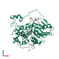
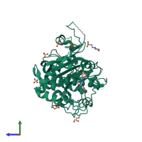
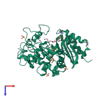
2zm7

X-ray diffraction
1.6Å resolution

Structure of 6-Aminohexanoate-dimer Hydrolase, S112A/G181D Mutant Complexed with 6-Aminohexanoate-dimer

Released:

Function and Biology Details

Reaction catalysed:
(N-(6-aminohexanoyl))(n) + H(2)O = (N-(6-aminohexanoyl))(n-1) + 6-aminohexanoate
Biochemical function:
  • not assigned
Biological process:
  • not assigned
Cellular component:
  • not assigned

Structure analysis Details

Assemblies composition:
homo dimer
monomeric (preferred)
PDBe Complex ID:
PDB-CPX-139359 (preferred)
Entry contents:
1 distinct polypeptide molecule
Macromolecule:
6-aminohexanoate-dimer hydrolase Chain: A
Molecule details ›
Chain: A
Length: 392 amino acids
Theoretical weight: 42.88 KDa
Source organism: Paenarthrobacter ureafaciens
Expression system: Escherichia coli
UniProt:
  • Canonical: P07061 (Residues: 1-28, 181-208; Coverage: 14%)
  • Canonical: P07062 (Residues: 29-180, 209-392; Coverage: 86%)
Gene names: nylB, nylB'
Sequence domains: Beta-lactamase
Structure domains:

Ligands and Environments

No modified residues

Experiments and Validation Details

Entry percentile scores
X-ray source: SPRING-8 BEAMLINE BL38B1
Spacegroup: P3221
Unit cell:
a: 96.684Å b: 96.684Å c: 113.164Å
α: 90° β: 90° γ: 120°
R-values:
R R work R free
0.172 0.172 0.187
Expression system: Escherichia coli