


Function and Biology Details
Biochemical function:
- not assigned
Biological process:
- not assigned
Cellular component:
- not assigned
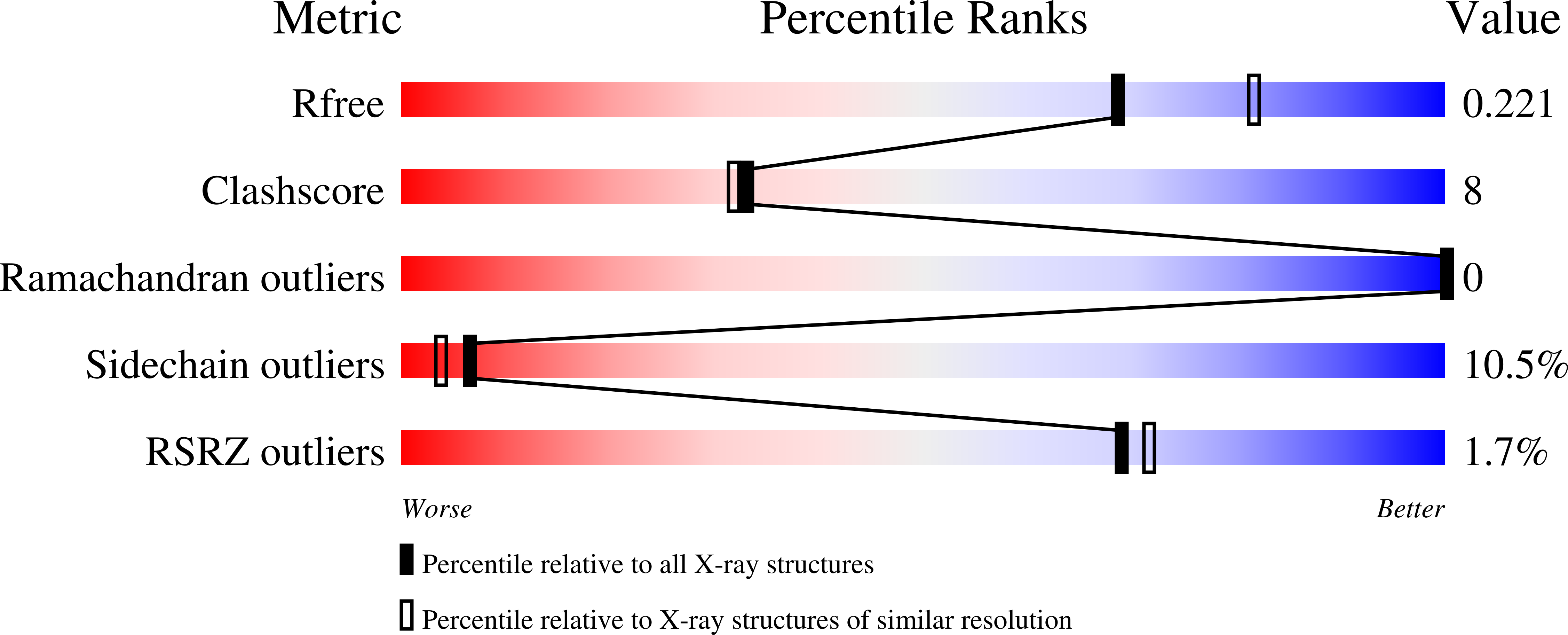
Structure analysis Details
Assembly composition:
homo dimer (preferred)
Assembly name:
PhiH1 repressor (preferred)
PDBe Complex ID:
PDB-CPX-178397 (preferred)
Entry contents:
1 distinct polypeptide molecule
Macromolecule:
PhiH1 repressor
Molecule details ›
Chains: A, B
Length: 111 amino acids
Theoretical weight: 12.49 KDa
Source organism: Haloarcula marismortui ATCC 43049
Expression system: Escherichia coli BL21(DE3)
UniProt:
Structure domains: Winged helix-like DNA-binding domain superfamily/Winged helix DNA-binding domain
Length: 111 amino acids
Theoretical weight: 12.49 KDa
Source organism: Haloarcula marismortui ATCC 43049
Expression system: Escherichia coli BL21(DE3)
UniProt:
- Canonical:
 Q5V043 (Residues: 1-108; Coverage: 100%)
Structure domains: Winged helix-like DNA-binding domain superfamily/Winged helix DNA-binding domain


