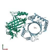
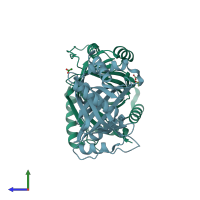
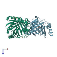
3bgy

X-ray diffraction
1.65Å resolution

Triclinic structure of Mimivirus Capping Enzyme Triphosphatase at 1.65 A

Released:
Entry authors: Smith P, Bennaroch D, Shuman S

Function and Biology Details

Reactions catalysed:
S-adenosyl-L-methionine + a 5'-(5'-triphosphoguanosine)-[mRNA] = S-adenosyl-L-homocysteine + a 5'-(N(7)-methyl 5'-triphosphoguanosine)-[mRNA]
GTP + a 5'-diphospho-[mRNA] = diphosphate + a 5'-(5'-triphosphoguanosine)-[mRNA]
A 5'-triphospho-[mRNA] + H(2)O = a 5'-diphospho-[mRNA] + phosphate
Biological process:
  • not assigned
Cellular component:
  • not assigned

Structure analysis Details

Assembly composition:
monomeric (preferred)
PDBe Complex ID:
PDB-CPX-178390 (preferred)
Entry contents:
1 distinct polypeptide molecule
Macromolecule:
Probable mRNA-capping enzyme Chains: A, B
Molecule details ›
Chains: A, B
Length: 237 amino acids
Theoretical weight: 27.56 KDa
Source organism: Acanthamoeba polyphaga mimivirus
Expression system: Escherichia coli
UniProt:
  • Canonical: Q5UQX1 (Residues: 1-237; Coverage: 20%)
Gene name: MIMI_R382
Structure domains: mRNA triphosphatase Cet1-like

Ligands and Environments

1 bound ligand:
No modified residues

Experiments and Validation Details

Entry percentile scores
X-ray source: NSLS BEAMLINE X29A
Spacegroup: P1
Unit cell:
a: 39.64Å b: 48.46Å c: 69.7Å
α: 74.62° β: 86.48° γ: 76.36°
R-values:
R R work R free
0.184 0.184 0.212
Expression system: Escherichia coli